
Ichiro Matsuo
Professor, Department of Applied Chemistry, Division of Materials and Environment, Graduate School of Science and Technology, Gunma University
Ichiro Matsuo received his M.Sc. degree in Science from Yokohama City University in 1992, under the supervision of Prof. Toru Sakakibara. From April 1992, he joined Meiji Dairies Corporation and was assigned to the Meiji Institute of Health Science, where he conducted research under the supervision of Dr. Katsumi Ajisaka. From October 1992-1993, he completed one-year contract research at the Laboratory for Synthetic Cellular Chemistry, RIKEN, under the supervision of Chief Scientist Dr. Tomoya Ogawa. In 1998, he obtained his Ph.D. in Agriculture from the Graduate School of Agricultural and Life Sciences, The University of Tokyo. Following a research period at the Laboratory of Microbiology, The University of Tokyo (under Prof. Katsuhiko Kitamoto), he served as a researcher at the Laboratory for Synthetic Cellular Chemistry, RIKEN (under Chief Scientist Yukishige Ito) starting in 2001. He was appointed Professor at the Graduate School of Engineering, Gunma University, in 2008. Since 2015, he has held the position of Professor at the Department of Molecular Science, Graduate School of Science and Technology, and has served in his current capacity since 2025.

Nozomi Ishii
Assistant Professor, Department of Applied Chemistry, Division of Materials and Environment, Graduate School of Science and Technology, Gunma University
She received her Ph.D. in Science and Technology from Gunma University in March 2018 under the direction of Professor Ichiro Matsuo. She then worked as a postdoctoral fellow at the University of Canterbury, New Zealand, from April 2018 to August 2020 under the supervision of Professor Antony J. Fairbanks. Since September 2020, she has been an Assistant Professor at Gunma University.
Endo-β-N-acetylglucosaminidase (ENGase) is an endoglycosidase that acts on the N,N’-diacetylchitobiose core of asparagine-linked glycans (N-glycans), cleaving the β-glycosidic linkage. ENGase is widely utilized as a research tool for glycan structural analysis because it releases N-linked glycans from glycoproteins. Additionally, ENGase is employed for glycoprotein glycan remodeling, with expanding applications in drug discovery research, making it an industrially valuable enzyme. To facilitate further ENGase studies, simple methods for detecting the glycoside hydrolase activity of ENGase are required. However, most existing activity assays require tedious manipulations. In this article, we introduce FRET (Förster resonance energy transfer)-quenching-based glycan molecular probes, developed through chemical glycan synthesis, that enable simple, highly sensitive, high-throughput, and real-time detection of ENGase activity and are suitable for inhibitor screening.
Since ENGase cleaves the chitobiose moiety of asparagine-linked (N-linked) glycans, glycoproteins, glycopeptides, or fluorescently labeled glycans can serve as substrates to detect its activity. When using glycoproteins as substrates such as RNase B, fetuin, or antibodies, ENGase acts on the protein, and the resulting change in molecular weight from glycan cleavage is analyzed by sodium dodecyl sulfate-polyacrylamide gel electrophoresis (SDS-PAGE). Substrate specificity can also be evaluated based on the reactivity toward these glycoproteins; however, quantitative analysis is difficult due to the inherent structural diversity of glycans on glycoproteins.
High-performance liquid chromatography (HPLC) or mass spectrometry methods using fluorescently labeled glycopeptides as substrates, which ensure structural homogeneity, allow for highly sensitive detection of enzymatic activity1-4. However, when using glycoproteins or glycopeptides as substrates, competition with peptide:N-glycanase (PNGase) can occur; therefore, when analyzing samples such as cytoplasmic fractions, it is necessary to ensure that only ENGase activity is detected.
In methods that detect ENGase activity via HPLC using a glycan library where the reducing ends of the glycans have been fluorescently labeled (e.g., via pyridylamination), substrate specificity can be inferred from differences in the profiles of the labeled glycans following ENGase treatment. The reducing ends of glycans labeled via reductive amination are in an open-ring form; they do not serve as substrates for PNGase, allowing specific detection of ENGase activity. Nevertheless, caution is required when evaluating substrate specificity, as some ENGases recognize the cyclic structure of the glycan's reducing end. In any case, detecting ENGase activity using these methods involves laborious procedures such as SDS-PAGE or HPLC, resulting in low throughput and an inability to monitor ENGase activity in real time.
Substrates for detecting exoglycosidase activity are commercially available as kits, allowing enzyme assays to be performed conveniently. However, kits for ENGase that enable simple activity detection were not commercially available. Therefore, we designed a chemically synthesized glycan molecular probe that generates fluorescence upon cleavage by ENGase, enabling simple, quantitative detection of ENGase glycoside hydrolase activity. Furthermore, using a microplate reader would enable high-throughput, real-time detection across multiple samples, thereby accelerating ENGase research, including the discovery of novel ENGases, the engineering of mutants with altered substrate specificity, and the screening of ENGase inhibitors.
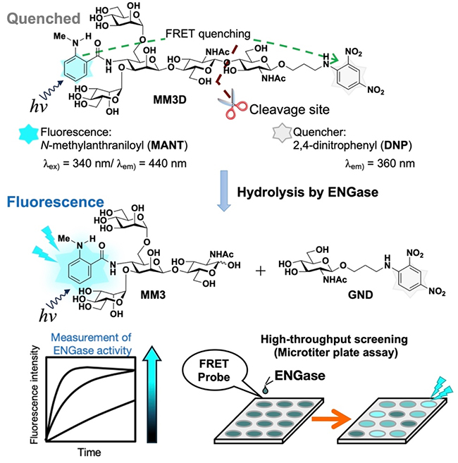
To this end, we developed a glycan molecular probe based on a Förster resonance energy transfer (FRET)-quenching system. FRET is a phenomenon in which excitation energy is transferred from a donor molecule to a nearby acceptor molecule. Thus, if a glycan molecule is chemically synthesized by introducing a fluorescent donor group and a quenching acceptor group in proximity within a glycan residue, no fluorescence is emitted (the FRET system remains dark) because energy from the excited fluorescent group is transferred to the quencher. Upon cleavage of the glycosidic bond by ENGase, intramolecular FRET quenching does not occur, and light from the fluorescent group is no longer quenched (the group continues to emit fluorescence). As a result, fluorescence intensity increases as the enzymatic reaction progresses, enabling real-time, highly sensitive detection of ENGase activity by monitoring this increase.
The concept of the FRET-quenched probe is shown in Figure 1. To minimize steric hindrance, we chose the N-methylanthraniloyl group (MANT) as the fluorophore and the 2,4-dinitrophenyl group (DNP) as the quencher. The underlying glycan structure was based on the conserved core pentasaccharide present in all asparagine-linked glycans. Regarding the positions of the functional groups across the chitobiose site cleaved by ENGase, the fluorophore was introduced at the non-reducing end, and the quencher was incorporated at the reducing end via an aminopropyl linker5.
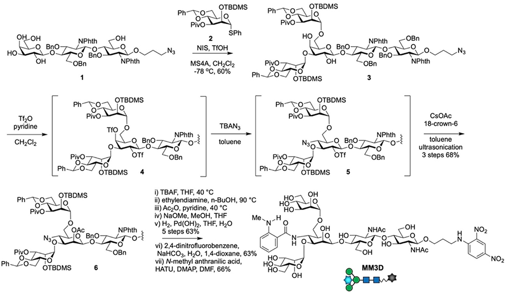
Scheme 1 shows the synthetic route to the FRET-quenched probe MM3D, which contains a pentasaccharide core structure. We constructed the β-mannose residue through the inversion of stereochemistry at the C-2 and C-4 hydroxy groups of a β-galactose residue6. This strategy was chosen because: 1) the high reactivity of the C-3 and C-6 hydroxy groups on the galactose residue allows the branched structure to be constructed in a single step; and 2) among the leaving groups (triflate groups, Tf) introduced to the C-2 and C-4 hydroxy groups of the galactose residue, the Tf group at C-4 exhibits higher reactivity, enabling selective introduction of an azide group at the C-4 position. Subsequent treatment with CsOAc and crown ether under ultrasonic conditions then leads to the desired β-mannose residue.
Specifically, using the reaction of trisaccharide acceptor 1 with mannosyl donor 2, the branched pentasaccharide skeleton was constructed in a single step to give pentasaccharide 3. Conversion of the C-2 and C-4 positions of the galactose residue in 3 to triflates afforded ditriflate 4. Subsequent reaction with tetrabutylammonium azide (TBAN3) in toluene at room temperature selectively introduced an azide group at C-4. After removing excess TBAN3 via extraction, treatment with CsOAc and 18-crown-6 ether under ultrasonic conditions yielded pentasaccharide 6 possessing the desired stereochemistry in 68% yield over 3 steps.
After the global deprotection of pentasaccharide 6 to yield a diamine intermediate, the difference in amino group reactivity was used to selectively introduce the DNP group at the reducing end, followed by the incorporation of the MANT group at the non-reducing terminal amino group, affording the pentasaccharide probe MM3D.
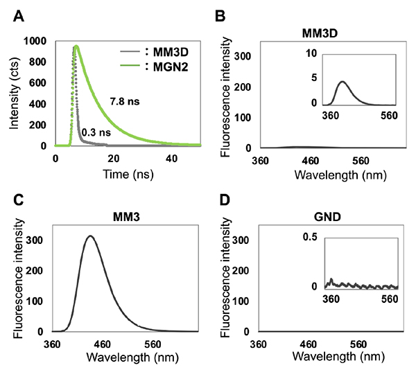
Next, we evaluated whether MM3D functions as a FRET-quenching glycan molecular probe by measuring the fluorescence lifetime of the MANT group. While the chitobiose derivative MGN2 (ManGlcNAc2) containing only the MANT group exhibited a fluorescence lifetime of 7.8 ns, the fluorescence lifetime of the MANT group on MM3D was 0.3 ns. This confirmed that the fluorescence of the MANT group was significantly quenched by the DNP group (Figure 2A). Furthermore, comparing the fluorescence intensity of MM3D with that of the tetrasaccharide MM3, which is the expected cleavage product generated upon complete hydrolysis of MM3D by ENGase, revealed that the fluorescence intensity of MM3 was approximately 50-fold higher than that of MM3D. This fluorescence enhancement was sufficient to track the enzymatic reaction (Figure 2B,C). Additionally, we confirmed that the DNP group in N-acetylglucosamine derivative GND emits no fluorescence (Figure 2D) and that no intermolecular quenching occurs among MM3D, MM3, and GND5.
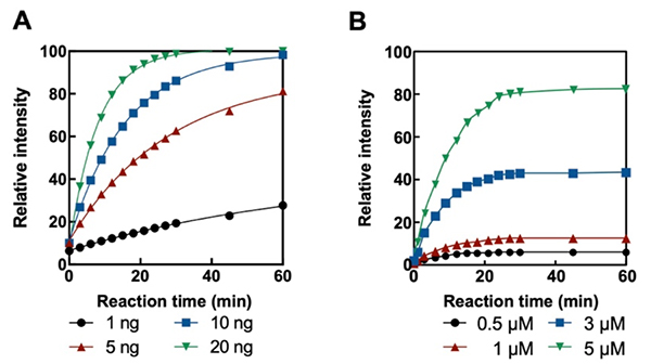
We next evaluated whether MM3D serves as a substrate for ENGase using Endo-M from Mucor hiemalis7. We fixed the MM3D concentration at 5 μM and varied the enzyme concentration, monitoring the reaction with a microplate reader at an excitation wavelength of 340 nm and an emission wavelength of 440 nm. At all enzyme concentrations, the fluorescence intensity at 440 nm increased over time compared to the initial state immediately after starting the reaction. Furthermore, because the rate of fluorescence increase was dependent on enzyme concentration, we concluded that the enzymatic reaction can be successfully monitored (Figure 3A).
HPLC analysis of the reaction mixture revealed that the peak corresponding to MM3D decreased with reaction time, accompanied by the appearance of peaks corresponding to chemically synthesized tetrasaccharide MM3 and monosaccharide GND. Subsequent Liquid chromatography mass spectrometry analysis detected molecular ion peaks corresponding to their calculated molecular weights, confirming that Endo-M cleaves the glycosidic linkage within the chitobiose core of MM3D. Furthermore, evaluating different probe concentrations demonstrated that the enzymatic reaction could be clearly tracked even at 0.5 μM, establishing that the assay can monitor the enzymatic reaction with high sensitivity and quantitativeness (Figure 3B).
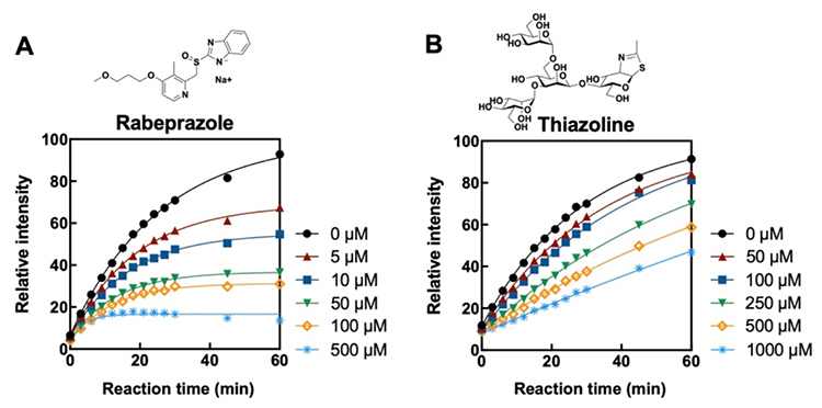
Next, because Endo-M activity was easily detected using a plate reader, we investigated whether MM3D could be used to evaluate ENGase inhibitors. Specifically, we assessed the Endo-M-catalyzed cleavage of MM3D in the presence of rabeprazole8, which inhibits human ENGase associated with NGLY1 gene deficiency (resulting in the loss of the N-glycanase 1 enzyme or its function), and thiazoline derivative9, a known ENGase inhibitor. Consequently, we observed a concentration-dependent deceleration of the enzymatic reaction with both rabeprazole and thiazoline, demonstrating that our MM3D-based ENGase evaluation platform can be used to screen novel inhibitors. Note that MM3D is currently commercially available from Tokyo Chemical Industry Co., Ltd. (TCI), allowing researchers to perform ENGase activity assays easily10.
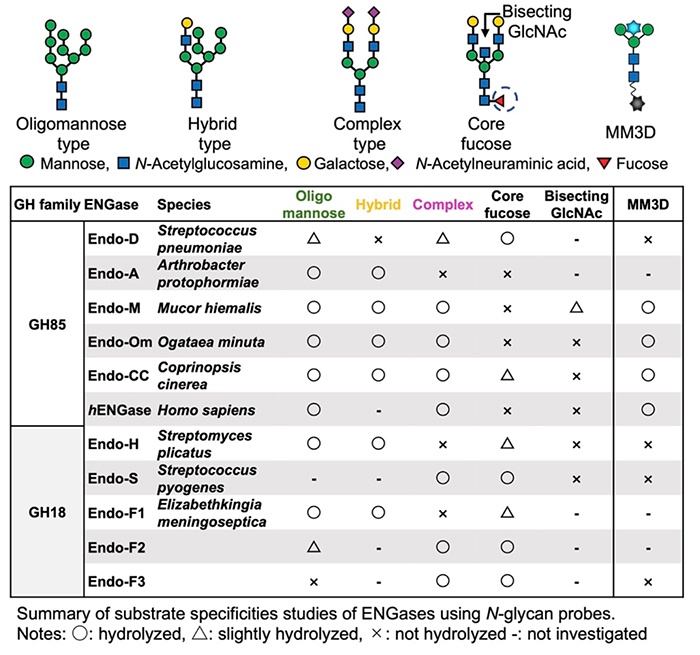
Since MM3D contains the conserved core structure of N-glycans targeted by all ENGases, we anticipated it would serve as a universal substrate for various ENGases. Therefore, we attempted to detect the enzymatic activities of ENGases from different origins using MM3D. We tested Endo-D from Streptococcus pneumoniae11, Endo-Om from Ogataea minuta12, Endo-CC from Coprinopsis cinerea13, and a human ENGase14, which are considered members of the GH85 family of glycoside hydrolases, as well as Endo-H from Streptomyces plicatus15, Endo-F3 from Flavobacterium meningosepticum16, and Endo-S from Streptococcus pyogenes17, which are regarded as GH18 family members. Table 1 summarizes the results of the enzyme reaction evaluations using MM3D, alongside previously reported substrate specificities for these ENGases. MM3D successfully detected activity for Endo-M, Endo-CC, Endo-Om, and the human ENGase, but not for Endo-D, Endo-H, Endo-S, or Endo-F3. Endo-D is known to act on the core pentasaccharide, and we confirmed that it also acts on the core pentasaccharide without the fluorescent group. Consequently, the failure of Endo-D to act on MM3D was caused by the fluorescent group introduced at the C-4 position of the β-mannose residue.
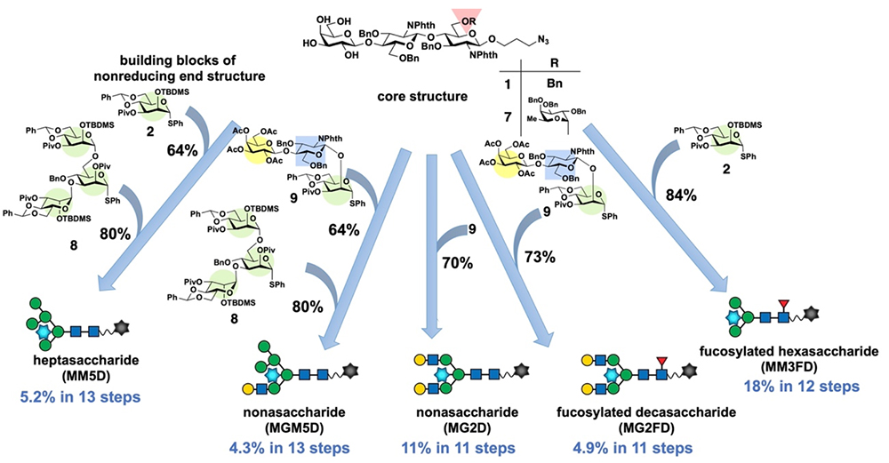
The pentasaccharide probe MM3D, with the conserved core structure of N-glycans, failed to detect the activity of ENGases that recognize specific glycan structures at the non-reducing end. Therefore, to detect the activity of various ENGases, glycan molecular probes with distinct non-reducing terminal glycan structures were required. To address this, we chemically synthesized probes bearing oligomannose, hybrid-type glycans, and complex-type glycans at the non-reducing end, as well as probes with a core fucose residue at the reducing end (Figure 5). These glycan skeletons were constructed in the same manner as those assembled by pentasaccharide probe synthesis. Specifically, taking advantage of the difference in reactivity among the hydroxy groups on the galactose residue of trisaccharide acceptor 1, the characteristic glycan structures at the non-reducing end were assembled via a sequential blockwise synthetic method. In the case of the heptasaccharide probe MM5D18, having an oligomannose-type glycan structure, the heptasaccharide skeleton was constructed by introducing mannosyl donor 2 at the C-3 position of the galactose residue of 1, followed by the introduction of branched mannotriose donor 8 at the C-6 position. For the hybrid-type probe MGM5D19, trisaccharide donor 9, which has a complex-type structure, was coupled at C-3 of acceptor 1, followed by reaction with the mannotriose donor 8 at C-6. In contrast, the biantennary complex-type nonasaccharide MG2D20 and the core-fucosylated complex-type decasaccharide MG2FD20 were constructed by introducing the trisaccharide donor 9 to trisaccharide acceptor 1 or tetrasaccharide acceptor 7 in a single step, giving the corresponding nonasaccharide and decasaccharide skeletons, respectively. Core fucosylated hexasaccharide MM3FD21 was assembled by reacting mannose donor 2 with tetrasaccharide acceptor 7 bearing the core-fucose structure. Conversion to glycans bearing the desired β-mannose residue was achieved through introduction of an azide group at the C-4 position and stereoinversion at the C-2 position via ditriflation at the C-2 and C-4 positions of the galactose moiety in these glycans. Finally, global deprotection and sequential introduction of the DNP and MANT groups yielded the target glycan molecular probes.
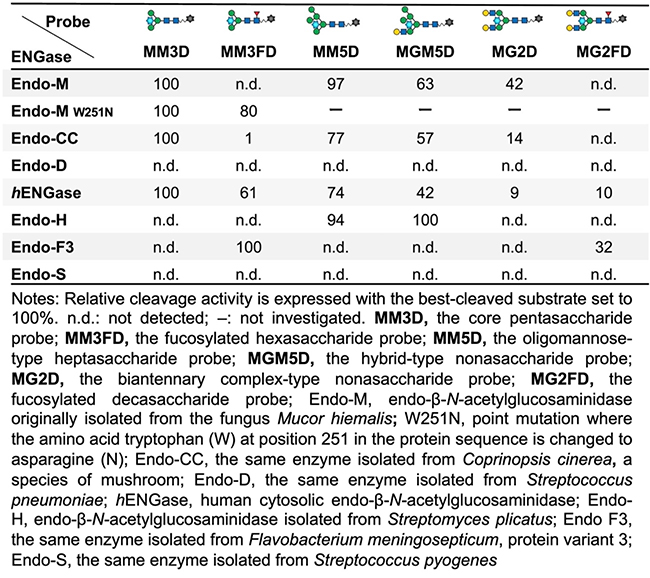
The activities of various ENGases were evaluated using six types of glycan molecular probes: the core pentasaccharide probe MM3D; the fucosylated hexasaccharide probe MM3FD; the oligomannose-type heptasaccharide probe MM5D; the hybrid-type nonasaccharide probe MGM5D; the biantennary complex-type nonasaccharide probe MG2D; and the fucosylated decasaccharide probe MG2FD. We elucidated the substrate specificity of each ENGase by reacting the same amount of enzyme with probes of different structures, each prepared at a fixed concentration. We evaluated the activity of commercially available ENGases, including Endo-M, Endo-CC, Endo-D, hEENGase, Endo-H, Endo-F3, and Endo-S. Specifically, we added each ENGase to 5 μM probe solutions and monitored the fluorescence intensities using a microplate reader. Table 2 shows the relative cleavage activities, with the activity against the probe substrate most efficiently cleaved by each enzyme set to 100%.
Endo-M efficiently cleaved the core pentasaccharide MM3D and the oligomannose-type heptasaccharide MM5D. However, its reactivity toward MGM5D and MG2D, which possess complex-type glycan structures at the C-3 position, was low. Meanwhile, it did not hydrolyze the fucosylated probes MM3FD and MG2FD. These results reflect the substrate specificity of Endo-M reported in previous studies22. Endo-M-W251N23 accepts the core-fucosylated hexasaccharides MM3FD and MM3D. Endo-CC showed a similar tendency to prefer oligomannose-type glycans over complex-type glycans. Furthermore, unlike Endo-M, Endo-CC was shown to slightly cleave the core-fucosylated hexasaccharide MM3FD. Kurogochi et al24. also reported the susceptibility of core-fucosylated glycans to cleavage by Endo-CC and our probe-based analysis reflected the substrate specificity of Endo-CC. No activity was detected for Endo-D with any of the glycan probes; as previously mentioned, this was likely due to the fluorescent group introduced at the bisecting position, which prevented proper assessment of its activity. hEENGase demonstrated low substrate specificity, hydrolyzing glycans independently of the structure at the non-reducing end. Hirayama et al. analyzed hEENGase activity using PA-labeled glycans but did not observe cleavage activity against core-fucosylated glycans1. However, we detected enzymatic activity against MM3FD and MG2FD; while further detailed analysis is required, this suggests that the cyclic structure at the reducing end of the GlcNAc residue plays an important role in hEENGase glycan recognition.
Next, we evaluated Endo-H, Endo-F3, and Endo-S, which belong to the GH18 family. The results showed that Endo-H acted on oligomannose-type and hybrid-type glycans, but not on complex-type glycans, core pentasaccharides, or fucosylated glycan probes. In contrast, Endo-F3 did not act on probes lacking a core fucose residue, indicating a strong recognition of the core fucose residue. Additionally, Endo-F3 preferred MM3FD over MG2FD. This indicates that Endo-F3 has a higher affinity for the glycan structure on the reducing-end side. Finally, we examined the activity of Endo-S, which is known to act specifically on antibody glycans. Endo-S showed no activity on any of the six probes. We attribute this to X-ray crystallographic analysis of Endo-S, which revealed a loop structure near the β-mannose residue25. We believe steric hindrance from the fluorescent group introduced to the substrate prevented recognition as a substrate.
We developed a FRET-based glycan molecular probe capable of simple, real-time detection of enzymatic reactions targeting ENGase. Regarding the chemical synthesis of the glycan probe, we established a synthetic route that significantly reduced the number of protection and deprotection steps by employing regioselective and stereoselective glycosylation of the four free hydroxy groups on the galactose residue. Furthermore, we efficiently synthesized the glycan molecular probe through a stereoinversion reaction converting a β-galactose residue into a β-mannose residue. We confirmed that our developed glycan molecular probe enables simple, real-time detection of ENGase activity across multiple samples using a microplate reader. Consequently, this method is suitable for comprehensive inhibitor screening. Suzuki and colleagues at RIKEN have demonstrated a link between hENGase and the rare disease NGLY1 deficiency26,27. We anticipate that our method will facilitate the discovery of novel ENGase inhibitors that could serve as therapeutic agents for NGLY1 deficiency.
Additionally, using glycan molecular probes with varying glycan structures has simplified the analysis of ENGase substrate specificity. Our probes enabled the rapid detection of activity and the analysis of substrate specificity for an unidentified ENGase. We hope that many ENGase researchers will actively utilize these glycan molecular probes, thereby contributing to the development of industrially useful ENGases, such as through the discovery of novel ENGases or the generation of ENGase mutants with altered substrate specificities via genetic engineering.
Finally, we confirmed that it is possible to detect the activity of enzymes such as Endo-S and Endo-D, whose activity was initially undetectable, by leveraging chemical synthesis techniques to optimize the site of fluorescent group attachment and the glycan structure. We aim to advance research that contributes to elucidating ENGase functions and conducting exploratory studies by utilizing glycan molecular probes accessible only through chemical synthesis, such as probes with tri- or tetraantennary complex-type glycans, probes with enhanced sensitivity achieved by modifying FRET pairs, and molecular probes capable of detecting intracellular ENGase activity.