Tadashi Suzuki
Chief Scientist, Glycometabolic Biochemistry Laboratory; RIKEN Pioneering Research Institute (PRI)
Dr. Suzuki obtained a Dr.Sci. (Doctor of Science) degree in 1997 from Department of Biochemistry and Biophysics, Graduate School of Science, University of Tokyo (Advisors: Prof. Yasuo Inoue/Assoc. Prof. Yasufumi Emori). From July 1997, he was a Post-doctoral Fellow/Research Scientist/Research Assistant Professor at State University of New York at Stony Brook (Prof. William J. Lennarz). In December 2001 he came back to Japan as a Researcher of the Precursory Research for Embryonic Science and Technology (PRESTO), Japan Science and Technology Corporation (JST). From February 2002 he also served as an RCF Assistant Professor at the Graduate School of Science, University of Tokyo, and in January 2004, he became Visiting Associate Professor at Osaka University Graduate School of Medicine. From October 2007, he was a Team Leader at Glycometabolome Team, Systems Glycobiology Research Group, RIKEN, and from April 2018 he holds a current position.
Endo-β-N-acetylglucosaminidase (ENGase) hydrolytically cleaves the glycosidic linkage in the N,N’-diacetylchitobiose moiety of the core region of N-linked glycans (N-glycans), and it has long been widely used as a tool for structural analysis of N-glycans. It has been suggested that cytosolic ENGases, which are widely distributed in animal cells including human cells, are involved in the degradation of free N-linked glycans (FNGs), but recently the ability of cytosolic ENGases to act directly on proteins has also been shown. While Engase-knockout (KO) mice show no particular phenotypes, deletion of the Engase gene can partially suppress embryonic lethality caused by gene knock-out of the Ngly1 gene, which encodes peptide:N-glycanase (PNGase), i.e., another cytosolic deglycosylating enzyme acting on N-glycans. ENGase is thus attracting attention as a potential drug target for NGLY1 deficiency, a rare human genetic disorder. Recently, part of the molecular mechanism by which ENGase suppresses the phenotype in Ngly1-KO mice has been clarified, revealing that it is quite complex, involving the E3 ubiquitin ligase complex SCFFBS2- ARIH1.
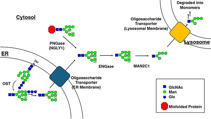
Historically, endo-β-N-acetylglucosaminidase (ENGase; EC 3.2.1.96) has been widely used to analyze the structure and function of asparagine-linked glycans (N-linked glycans)1. Mammalian ENGase activity was first reported in 1974 by the laboratory of Professor Akira Kobata2, and its localization mainly in cytosol was first reported by the group of Professor Jean Montreuil in France3. In 1981, ENGase activity was first reported in humans4,5. According to the classification in the CAZy database6, ENGases are classified into the glycoside hydrolase (GH) families 18 and 85, and ENGases widely found in the cytosol of animal cells belong to GH85. Mammalian ENGases are involved in the processing of free N-glycans (FNGs) in the cytosol ("non-canonical glycan metabolism") (Figure 1). If the ENGase is deficient, the subsequent action of cytosolic α-mannosidase (Man2C1) is also inhibited, thereby resulting in the accumulation of unprocessed glycans, i.e., Man8-9GlcNAc27.
The human Engase gene was reported in 200210. This enzyme acts preferably on high-mannose type glycans, but hardly at all on glycans with core fucose (Fuc)5,11. This substrate specificity, however, needs to be carefully re-examined, since glycoproteomic analyses from mammalian samples often identify HexNAc-dHex (presumably GlcNAc-Fuc)12-14, which can be assumed to be formed by the action of ENGase, as a glycan on proteins. In the future, it will be necessary to investigate how the presence or absence of core Fuc affects human ENGase activity using, for example, chemically synthesized glycopeptides containing core Fuc15.
Human ENGase exhibits a very broad tissue distribution10, and therefore was predicted to be involved in certain fundamental cellular functions. However, its precise functional importance remains unclear, partly because no significant phenotype was observed for Engase-KO mice16. It is noteworthy that, while the structural profile of FNGs in the cytosol of Engase-KO mouse embryonic fibroblasts changed significantly, the total amount of FNGs did not change significantly7. This result suggests that the cytosolic glycan catabolism may also occur via an alternative pathway (such as the autophagy-lysosome pathway) that targets cytosolic FNGs to lysosomes, in an ENGase-Man2C1 processing (Figure 1)-independent fashion. Alternatively, although not clarified in any organisms, the release of FNGs into extracellular space may be possible under certain conditions.
Surprisingly, Engase-KO partially suppressed the embryonic lethality of Ngly1 (PNGase)-KO mice16; PNGase/NGLY1 is another cytosolic deglycosylation enzyme acting on N-glycans. As mentioned earlier, Engase-KO mice appear quite normal, similar to wild-type, and therefore there is some expectation that the specific inhibitors for ENGase could serve as a viable therapeutic option for individuals with NGLY1 deficiency. We have proposed that NGLY1 deficiency could be due to upregulation of N-GlcNAc-containing proteins formed by ENGase that directly act on cytosolic misfolded glycoproteins (“N-GlcNAc hypothesis”)17,18.
More recently, Dr. Yukiko Yoshida of the Tokyo Metropolitan Institute of Medical Science and others have elucidated one of the precise roles of ENGase in the pathogenesis of NGLY1 deficiency19. The mechanism is very complicated. For details, please refer to Dr. Yoshida's Glycoforum article (https://www.glycoforum.gr.jp/article/28A13.html). In summary, “Especially in the NGLY1 deficiency state, N-linked glycans on the transcription factor NRF1/NFE2L1 are partially removed by ENGase Note 1) (this ‘partial’ is critical; otherwise NRF1 would not be recognized by the glycan-recognizing E3 ubiquitin ligase complex, SCFFBS2-ARIH1). Then, the hydroxyl group (mainly at position 6) of the resulting N-GlcNAc becomes an excellent acceptor for atypical ubiquitination by SCFFBS2-ARIH1, and the subsequent accumulation of NRF1 with atypical ubiquitin chains accounts for, at least in part, the pathogenesis of NGLY1 deficiency" (you see, it is indeed very complicated.).
What is important here is that the "ubiquitination status of NRF1" predicted here corresponds very well to the mouse phenotypes we observed (Figure 2).
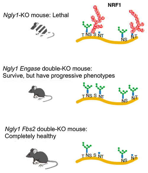
For example, (Top) In Ngly1-KO mice, the accumulation of ubiquitinated NRF1 has pathological consequences (for C57BL/6 mice, it results in embryonic lethality16). (Middle) In Ngly1 Engase-double KO mice, ENGase does not function even in the absence of Ngly1, so an excellent acceptor for SCFFBS2-ARIH1-mediated ubiquitination (N-GlcNAc) is not formed, and accordingly, non-canonical ubiquitination does not occur. On the other hand, since SCFFBS2-ARIH1 can also induce ubiquitination of other glycoproteins, noncanonical ubiquitination by SCFFBS2-ARIH1 is not completely inhibited. In C57BL/6 mice, embryonic lethality of Ngly1 Engase-double KO mice is partially rescued, but the resulting mice exhibit phenotypes similar to individuals with symptoms of NGLY1 deficiency, for example, smaller stature or development of tremors/bent spine as they age16. (Bottom) In Ngly1 Fbs2-double KO mice, since there is no recognition of N-glycans by FBS2 in the first place, ubiquitination by SCFFBS2-ARIH1 does not occur at all. Accordingly, in C57BL/6 mice, the Ngly1 Fbs2-double KO almost completely suppresses the embryonic lethality of Ngly1-KO, and motor function in the offspring of Ngly1 Fbs2-double KO mice is comparable to that of wild-type mice, and no phenotypic changes associated with aging have so far been observed20.
Here, we must consider the unique structural characteristics of NRF1. NRF1 has its N-linked glycosylation sites concentrated in a central region (called “the NST domain”), and there are no Lys residues, which typically serve as ubiquitination acceptor sites, around it (Figure 3). This is likely why NRF1 undergoes atypical ubiquitination via SCFFBS2-ARIH1. However, the question remains whether other substrates undergo similar N-GlcNAc-mediated atypical ubiquitination. Cellular-level analysis suggests that the lethality of FBS2 overexpression in Ngly1-KO HeLa cells is mitigated by simultaneous deletion of NRF120, implying that NRF1 is the primary acceptor for atypical ubiquitination that causes HeLa cell lethality. However, whether other proteins undergo similar N-linked glycosylation-mediated atypical ubiquitination, and if so, what impact they have on the pathogenesis of NGLY1 deficiency, remain unresolved and are subjects for future research.
Note 1) Regarding the significance of the ENGase reaction, it is also speculated that by "partially" removing the glycan chain, it will facilitate access to NRF1 by the SCFFBS2-ARIH1 complex, but this is way too complicated to discuss in detail in the main text.

As mentioned earlier, no significant phenotypes have been observed in Engase-KO mice so far, and the functional importance of mammalian ENGase remains a mystery. However, I believe that it should play an important role in some context, and I look forward to further research in this area.
Finally, a word related to this review series: Does human ENGase have glycosynthase activity? I myself have not noted any research indicating that it does, but the sequence around the active site, including residues considered important for glycosynthase activity near the active site21, is conserved between human ENGase and the C. elegans ENGase orthologue, and since glycosynthase activity is observed in the C. elegans worm’s ENGase22, I would probably say “yes” to this question.
I gratefully acknowledge Drs. Yukiko Yoshida (Tokyo Metropolitan Institute of Medical Science) and Chengcheng Huang (iGCORE, Nagoya University) for critical reading of the manuscript.