
Tomio Yabe
Professor, Faculty of Applied Biological Sciences, Gifu University
Dr. Yabe completed the doctoral program in Agricultural Chemistry at the Graduate School of Agricultural Science, Tohoku University, in 1999. He subsequently worked as a resident at Nippon Roche K.K., a postdoctoral fellow at the Massachusetts Institute of Technology, a visiting researcher at Harvard University, and a postdoctoral fellow at the Tokyo Metropolitan Institute for Neuroscience.
In 2004, he joined the Faculty of Applied Biological Sciences at Gifu University as an Assistant Professor. He was promoted to Associate Professor in 2007 and has served as Professor since 2016.
His research interests include functional food biochemistry and carbohydrate biochemistry. He received the Japan Association for Dietary Fiber Research Award in 2021.
Dietary fiber is known to exert a variety of physiological functions, including promoting bowel regularity and suppressing postprandial rises in blood glucose and serum cholesterol; many of these functions have been attributed to short-chain fatty acids produced by gut microbiota fermentation or to the physicochemical effects of indigestibility. Pectin, a water-soluble dietary fiber, is not degraded by human digestive enzymes and can therefore come into direct contact with the small-intestinal epithelium before it is fermented in the large intestine. In recent years, pectin has been found to alter the morphology of small-intestinal villi and to interact in a structure-specific manner with the extracellular matrix and cell-surface molecules of epithelial cells. In this review, from the viewpoint of the homogalacturonan backbone and the rhamnogalacturonan-I side chains—two distinct structural domains that act on distinct target molecules, I summarize the current knowledge about interactions between pectin and intestinal epithelial cells, and propose a working hypothesis that pectin acts as a form of “chemical information” recognized by epithelial cells.
Dietary fiber possesses a wide range of physiological functions that contribute to the maintenance of health by not only improving bowel movements but also suppressing rises in serum cholesterol and postprandial blood glucose levels and modulating immune function1,2. Many of these functions have been explained by the actions of short-chain fatty acids produced through fermentation by the gut microbiota residing in the large intestine, or by the physicochemical effects of dietary fiber, such as water retention, viscosity, and adsorption, which arise from its indigestible nature1. At the same time, the existence of a pathway by which dietary fiber itself acts directly on the gastrointestinal epithelium—one that cannot be fully explained by these two mechanisms alone—has also been pointed out.
Pectin, a type of water-soluble dietary fiber, is abundant in fruits and vegetables and is one of the major polysaccharides constituting the cell walls of terrestrial plants. Its basic backbone is homogalacturonan (HG), a chain of α-1,4-linked D-galacturonic acid in which some of the carboxy groups are methyl-esterified3,4. In addition, pectin contains rhamnogalacturonan-I (RG-I), in which neutral-sugar side chains such as galactan, arabinan, and arabinogalactan are attached to a backbone of alternating D-galacturonic acid and L-rhamnose, as well as the highly branched rhamnogalacturonan-II (RG-II)3,4 (Figure 1). The structure of pectin varies greatly across plant species, tissues, degrees of ripeness, and extraction methods, making it difficult to elucidate structure–function relationships5. For details of each structure, the reader is referred to excellent reviews3-5.
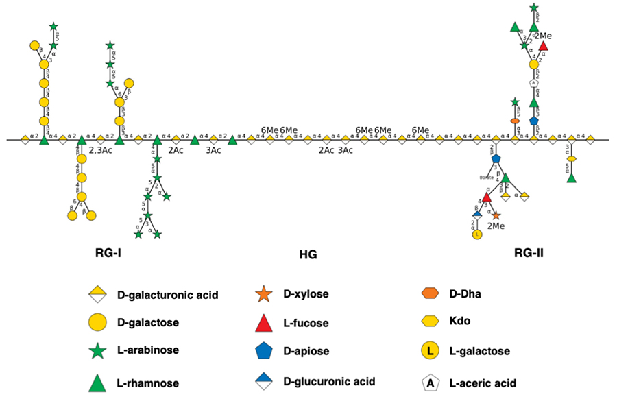
Pectin is not degraded by human digestive enzymes; it passes through the small intestine and reaches the large intestine, where it is almost completely fermented by the gut microbiota6. However, because substantial fermentation of pectin occurs mainly in the part of the small intestine beyond the terminal ileum, pectin can come into direct contact with the small-intestinal epithelium before substantial fermentation begins7. Indeed, it has long been reported that ingestion of pectin induces morphological changes, such as elongation of small-intestinal villi and branching of crypts8,9, and that these changes occur even under germ-free conditions9, supporting the hypothesis that a direct pathway exists independent of fermentation. Furthermore, there are reports that pectin binds to cell-surface lectin receptors and pattern-recognition receptors associated with the gastrointestinal tract, exerting anti-inflammatory and immunomodulatory actions7,10,11, and its possible role as a “ligand” for epithelial cell molecules has been discussed.
Nevertheless, for a long time, it remained unclear which molecules in or on small-intestinal epithelial cells pectin acts on, which structural moieties of pectin mediate these interactions, and what the underlying molecular mechanisms and physiological significance of these actions are. We have focused on the interaction between the intestinal epithelium and water-soluble dietary fibers that are not absorbed by intestinal villi, and have investigated their mode of action. In this review, I first outline the phenomenon whereby pectin alters the morphology of the small-intestinal epithelium and its molecular mechanism (Sections 2 and 3), and then describe the effect of HG, the backbone of pectin, on the interaction between the extracellular matrix protein fibronectin (FN) and integrin β1 (Section 4). I consider these findings from the perspective of structural specificity—namely, that the HG backbone and the RG-I side chains, two distinct structural domains, act on distinct epithelial molecules. Then, I incorporate these findings into a working hypothesis (Section 5).
It has long been known that pectin induces morphological changes in the small intestine. When rodents are given pectin at relatively high doses (on the order of 1–10% of the diet), elongation of small-intestinal villi and branching and proliferation of crypts are observed8,9,12,13. The small-intestinal epithelium is a tissue that is continuously renewed every several days: intestinal stem cells (ISCs) at the crypt base self-renew while giving rise to daughter cells, which proliferate and differentiate as they migrate toward the villus tip14. Villus elongation is therefore understood as a consequence of a change in the balance between proliferation and differentiation of stem and progenitor cells. Because such pectin-induced morphological changes occur even under germ-free conditions9, and because direct contact with cultured intestinal epithelial cells promotes cell proliferation (Section 3), pectin is considered to act directly on the epithelium itself.
The question that arises here is which structural moiety of pectin is responsible for this action. We found that the arabinogalactan moieties contained in the RG-I side chains of pectin from persimmon (Diospyros kaki) are involved in the interaction with small-intestinal epithelial cells15. That is, the candidate active sites responsible for morphological change are the neutral-sugar side chains rather than the HG backbone.
To examine this point in vivo, we first gave persimmon-derived pectin to senescence-accelerated mouse prone 8 (SAMP8) mice, an accelerated-aging model16. As a result, although villus length itself did not change markedly in aged SAMP8 mice, the pectin diet caused slight (10%) elongation of the villi in the middle part of the small intestine. More evident was the effect on stem-cell activity: the pectin-diet group showed increased expression of the Paneth-cell marker Lyz1 and of Olfm4, a marker of actively proliferating ISCs, and increased number of lobes in intestinal organoids prepared from excised crypts. These findings suggest that pectin enhances the regenerative capacity of ISCs16,17. In this study, however, it was also revealed that aging of SAMP8 mice is not itself accompanied by malabsorption of nutrients, so that the model is not necessarily suitable as a model of age-associated malabsorption16.
Next, to evaluate the physiological significance of the side chains more directly, we administered, via drinking water, Yuzu (Citrus junos) pectin side chains (the RG-I fraction prepared by digesting the backbone with pectinase) to mice in which mild malnutrition had been induced with a low-protein diet (protein-deficient; PD)18. As a result, the Yuzu pectin side chains (high-dose group) significantly elongated the jejunal villi, demonstrating for the first time that pectin side chains can cause villus elongation in vivo under PD conditions. On the other hand, the weight loss itself was not reversed, and the malnutrition was not completely ameliorated (partial efficacy). Gene expression analysis showed that the Yuzu pectin side chains reduced the expression of the duodenal glucose transporter Glut2 and the sucrase–isomaltase complex Si, indicating a tendency to suppress carbohydrate utilization. They further suppressed the hepatic lipid accumulation that had been elevated under PD. These results indicate that pectin side chains can reorganize the pattern of nutrient absorption by altering villus morphology, and that villus elongation and the absorption of individual nutrients do not necessarily coincide18.
Taken together, these findings suggest that the RG-I side chains of pectin may act as the active sites influencing small-intestinal epithelial morphology and ISC activity, and that this action may be meaningful in physiological contexts such as the prevention of malnutrition in the elderly19. However, these are all observations at the phenomenological level, and the molecular mechanism—how the information carried by the side chains is received by epithelial cells, conveyed to stem cells, and converted to stem-cell proliferation—cannot be explained by these alone. In the next section, I describe the in vitro evidence for this mechanism.
To understand the phenomena of the previous section at the molecular level, we used Caco-2 cells derived from human colon cancer. When differentiated, Caco-2 cells exhibit morphology and functions resembling those of the absorptive epithelial cells of the small intestine, and are therefore widely used as an in vitro model of the small-intestinal epithelium. Our focus was on cell-surface heparan sulfate (HS). HS is a sulfated polysaccharide, a glycosaminoglycan present on the surface of all animal cells and in the extracellular matrix; it forms various structures depending on its sulfation pattern and acts as a co-receptor for ligands such as Wnt and fibroblast growth factor20. In the small intestine, HS on the epithelial cell surface is also known to regulate crypt homeostasis through Wnt signaling21.
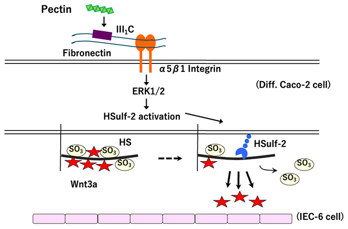
We first found that exposing differentiated Caco-2 cells to prune pectin (Prunus domestica L.) markedly altered the disaccharide composition (sulfation structure) of cell-surface HS22. Specifically, the proportion of non-sulfated disaccharides increased, and that of disaccharides bearing 6-O-sulfate groups decreased. This change was accompanied by increased expression of human HS 6-O-endosulfatase-2 (HSulf-2), the enzyme that removes 6-O-sulfate groups from HS at the cell surface, and by decreased expression of HSulf-1. Furthermore, pre-mixing pectin with the FN III1-C fragment (FN-III1C) or pretreating the cells with the arginine-glycine-aspartic acid (RGD) peptide suppressed the induction of HSulf-2. In addition, pectin stimulation transiently phosphorylated the extracellular signal-regulated kinase ERK1/2 (maximal at 5 min after stimulation), and an ERK1/2 inhibitor suppressed the induction of HSulf-2. These results suggest that pectin activates ERK1/2 via FN and α5β1 integrin and alters the sulfation structure of HS through the expression of HSulf-222. In this study, a preliminary finding that pectin binds to FN-III1C was also obtained; the molecular basis of this direct binding is described in Section 4.
Next, we examined the biological significance of this change in HS structure using a co-culture system of differentiated Caco-2 cells (an epithelial model) and rat IEC-6 cells (a crypt-cell model)23. Pectin-treated Caco-2 cells secreted some growth factors toward the basolateral side and promoted the proliferation of IEC-6 cells in a concentration-dependent manner. Whereas pectin increased the expression of Wnt3a (mRNA and protein) in Caco-2 cells, the structurally altered HS exhibited markedly reduced binding to Wnt3a. That is, pectin is thought to promote the secretion of Wnt3a from Caco-2 cells by altering the sulfation pattern of HS, thereby reducing the affinity between HS and Wnt3a. Wnt3a is secreted from cells such as Paneth cells, which form the stem-cell niche in the crypt, and is a factor that supports the proliferation of stem and progenitor cells24. The idea that 6-O-desulfation of HS weakens binding to Wnt and thereby regulates signaling is consistent with findings in other systems 25.
Taken together, these findings suggest that pectin-induced alteration of small-intestinal villus morphology may be explained by the pathway “pectin → FN/α5β1 integrin → ERK1/2 → 6-O-desulfation of HS by HSulf-2 → release of Wnt3a → proliferation of crypt cells” (Figure 2). This is a working hypothesis that may, at the molecular level, connect pectin to the in vivo villus elongation and enhanced ISC activity described in Section 2. On the other hand, it should be noted that these are in vitro findings using colon cancer–derived cells, and that the involvement of FN and integrin was inferred mainly from inhibition experiments and preliminary binding data. In the next section, therefore, I describe the results of intermolecular interaction analyses conducted to directly examine how pectin binds to FN and how this binding affects the interaction between FN and integrin.
As described in the previous section, the involvement of FN and α5β1 integrin in the action of pectin was suggested, but this inference was based mainly on inhibition experiments and preliminary binding data. FN is a large extracellular matrix glycoprotein formed by a succession of three types of modules—type I, type II, and type III—and plays a central role as a scaffold for cell adhesion, migration, and proliferation26. Cells adhere to FN by recognizing, via integrin α5β1, the RGD sequence on the III10 module of FN, with binding enhanced by the synergy sequence present on the III9 module of FN27. We therefore conducted intermolecular interaction analyses to directly determine where pectin binds to FN and how this binding affects the interaction between FN and integrin28.
The key here is to evaluate the structure of pectin by examining its component parts. From commercial pectin, we prepared four water-soluble pectins (WSPs) differing in structure28: Citrus WSP, consisting of a methyl-esterified HG backbone that contains approximately 88% uronic acid and almost no neutral sugars; pectic acid obtained by demethyl-esterifying Citrus WSP (PE Citrus WSP); and Kaki WSP from persimmon and Yuzu WSP from yuzu, both obtained by enzymatically digesting the backbone to enrich the neutral-sugar side chains (RG-I). Surface plasmon resonance (SPR) analysis of the binding of these WSPs to FN fragments revealed that the binding of the type III module of FN (FN-III1C) to Citrus WSP, composed of the methyl-esterified HG backbone, was clearly detectable (with a dissociation constant KD on the order of 10-7–10-8 M), and to the RG-I side chains of Kaki and Yuzu WSPs, weakly or hardly detectable. Furthermore, demethyl-esterification of Citrus WSP reduced its binding to FN. These results indicate that the binding is structurally specific in that among pectins, the methyl-esterified HG backbone binds preferentially to the FN type III module. Because it also attaches to an FN fragment containing the cell-adhesion domain, Citrus WSP is considered to bind to multiple sites of the FN type III module.
Next, we investigated the effect of this binding on the interaction between FN and integrin. Citrus WSP bound only weakly to integrin α5β1 itself. On the other hand, in an ELISA system in which an integrin β1–mimicking probe (the anti-FN antibody HFN7.1, which recognizes the cell-adhesion domain of FN-III10) was bound to plate-adsorbed FN, Citrus WSP reduced the binding of the antibody in a concentration-dependent manner28. Curve analysis showed that only the coefficient corresponding to the maximal binding capacity decreased, with no change in binding affinity; therefore, pectin was interpreted not as competitively blocking the RGD sequence but as substantially reducing the number of exposed cell-adhesion sites on FN. A similar perturbation was observed with low-methyl-esterified polygalacturonic acid and heparin, and was more pronounced for adsorbed FN than for FN in solution.
From these results, the methyl-esterified HG backbone is thought to bind to the type III module of FN, probably through hydrophobic interactions, and to perturb FN–integrin β1 binding by altering the structure of FN and the exposure of its cell-adhesion sites (in the demethyl-esterified form, the exposed carboxyl groups may bind electrostatically to the heparin-binding site). Because pectin—particularly the low-methyl-esterified form—can permeate the mucus layer and make contact with epithelial cells29, such perturbation could also occur in vivo. What is important here is that, in contrast to the RG-I side chains involved in the morphological change (Sections 2 and 3), the HG backbone acts on the FN–integrin axis, indicating that different structural domains of pectin target distinct epithelial molecules. The “perturbation of FN–integrin binding” in this section may appear, at first sight, to contradict the finding described in Section 3 that “pectin activates signaling via FN and integrin.” This apparent contradiction is discussed in the next section from the perspective of structural specificity.
The findings outlined in this review can be brought together into a single cohesive narrative: “different structural domains of pectin target different molecules of the small-intestinal epithelium.” The neutral-sugar side chains of RG-I (arabinan, galactan, and arabinogalactan) are involved in the morphological change of small-intestinal villi and in the activity of intestinal stem cells (Section 2), and their action may be explained by the modification of the sulfation structure of HS via FN, α5β1 integrin, and ERK1/2, and the accompanying release of Wnt3a (Section 3). The HG backbone, on the other hand—particularly the methyl-esterified backbone—binds to the type III module of FN and perturbs FN–integrin β1 binding (Section 4). Physiological functions dependent on side-chain structure are not limited to mechanisms of morphological change; for example, it has been reported that the amelioration of experimental colitis by pectin occurs in a manner dependent on the neutral-sugar side chains30, supporting the view of “different functions for different structural domains.”
There is, however, a point in this integrated narrative that must be dealt with directly. Whereas the mechanism in Section 3 suggests that pectin activates signaling via FN and integrin, Section 4 shows that the HG backbone perturbs FN–integrin β1 binding; the two appear, at first sight, to contradict each other. Several possible reasons for this apparent discrepancy can be considered. First is the difference in the structure of the pectin used (a purified fraction from prunes in Section 3, versus structurally defined Citrus HG in Section 4). Second is the difference in the state of FN: the perturbing action is more pronounced when FN is adsorbed to a matrix than when it is in solution, and in vivo FN exists in both the soluble form and the matrix-adsorbed form. Third, the very phenomena being observed differ (induction of intracellular signaling versus antibody/integrin binding). Conceivably, the binding of pectin, by changing the structure of FN or state of FN assembly, has a dual role—impeding adhesion in some contexts and triggering signaling in others. At present, these cannot be unified into a single mechanism, and this remains a subject for future verification.
A more fundamental question is where in the body these interactions take place. Because FN and integrin are normally localized on the basolateral side of epithelial cells, it is not yet clear whether pectin passing along the luminal side can contact them—that is, whether functional FN and integrin exist on the luminal side. In addition, it will be necessary to identify the minimal active unit of each domain, to demonstrate the molecular mechanism directly in vivo rather than in cellular models, and to clarify whether the actions of the two domains are independent or cooperative. It also remains necessary to keep in mind that much of the mechanism presented in this review is based on in vitro findings and inferences using human colon cancer–derived cells.
On the basis of these reservations, we wish to propose the following working hypothesis: although pectin is a food component that is not absorbed as a nutrient, its distinct structural domains act as a form of “chemical information” recognized by small-intestinal epithelial cells and thereby regulate epithelial homeostasis. Specifically, the HG backbone acting on the FN–integrin axis, a key regulator of cell adhesion, whereas the RG-I side chains affect villus morphology and stem-cell activity. If this view is correct, structurally defined pectin may be expected to provide design principles for functional food materials aimed at preventing malnutrition in the elderly and at controlling the intestinal barrier and epithelial regeneration. Indeed, attempts to guide cellular responses by controlling the binding specificity of FN and integrin have also been proposed31; an understanding of the structure-specific interactions of pectin will help to answer, in molecular terms, the questions of “why and how” we should consume dietary fiber.