Shinji Miyata
Associate Professor, Faculty of Agriculture, Tokyo University of Agriculture and Technology
After training under Professor Ken Kitajima at Nagoya University, Shinji Miyata received his Ph.D. in 2006. He then worked as a postdoctoral fellow under Professor Victor D. Vacquier at the University of California, San Diego. In 2007, he began research on glycosaminoglycans as a postdoctoral fellow under Professor Hiroshi Kitagawa at Kobe Pharmaceutical University. After serving as a designated assistant professor at Nagoya University from 2013 to 2019, he established his laboratory at Tokyo University of Agriculture and Technology in 2019. His research focuses on the functions of hyaluronan-proteoglycan complexes in the nervous system.

Hiroshi Kitagawa
Professor and President, Laboratory of Biochemistry, Kobe Pharmaceutical University
Hiroshi Kitagawa received his Ph.D. in Biochemistry in 1991 from Kyoto University. He then worked as a postdoctoral fellow at Cytel Corporation under Dr. James C. Paulson. In 1994 he obtained an assistant professor position at the Department of Biochemistry, Kobe Pharmaceutical University. He was promoted to associate professor in 2000 and full professor in 2005. He has served as president since 2022. He received the young scientist award of the Japanese Society of Carbohydrate Research in 1999, the PSJ (Pharmaceutical Society of Japan) award for young scientists in 2001, the young investigator award of the Japanese Biochemical Society in 2002, and the PSJ award for Divisional Scientific Promotions in 2013. His research focuses on the function and regulation of the biosynthesis and degradation of sulfated glycosaminoglycans to clarify the causes of various disorders.
Proteoglycans are glycoconjugates in which specific glycan chains are attached to core proteins, and they regulate diverse biological processes at the cell surface and in the extracellular matrix. Recent studies have clarified the molecular mechanisms involved in the initiation, elongation, sulfation, and termination of glycan biosynthesis on proteoglycans. This article provides an overview of proteoglycan biosynthesis in mammalian cells, with emphasis on recent advances.
Animal tissues consist of cells and the extracellular matrix that surrounds them. The extracellular matrix is not merely a scaffold that supports cells, but a dynamic environment that regulates cell proliferation, differentiation, migration, adhesion, and signaling responses. Proteoglycans are one of the major classes of molecules that constitute the extracellular matrix and cell surface1. Proteoglycans are defined as molecules in which one or more glycosaminoglycan (GAG) chains are covalently attached to a core protein1. Several classes of GAG chains exist, depending on their constituent sugars. This article focuses on two representative GAG chains: chondroitin sulfate (CS) and heparan sulfate (HS).
CS is a linear polysaccharide composed of repeating disaccharide units of [-4GlcAβ1-3GalNAcβ1-]n2. In contrast, HS consists of repeating disaccharide units of [-4GlcAβ1-4GlcNAcα1-]n3. These GAG chains are elongated through a common linkage tetrasaccharide formed on specific Ser residues of core proteins. When a CS chain is elongated, the resulting molecule is a CS proteoglycan; when an HS chain is elongated, the resulting molecule is an HS proteoglycan.
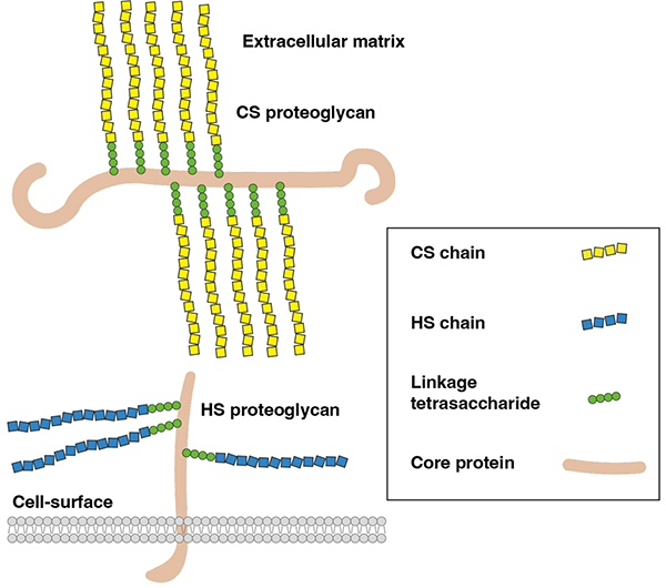
Representative CS proteoglycans include aggrecan, versican, neurocan, and brevican, which are secreted extracellular molecules that contribute to extracellular matrix formation4 (Figure 1). Representative HS proteoglycans include syndecans and glypicans, which localize to the cell surface and regulate the functions of growth factors and adhesion molecules1,3. In rare cases, a single core protein can carry both CS and HS chains; however, CS and HS do not coexist within a single GAG chain.
During biosynthesis, the linear repeating disaccharide backbone undergoes sulfation and epimerization, thereby generating highly diverse structures. The position and degree of sulfation and epimerization are thought to generate specific domain structures within individual GAG chains, allowing them to interact with defined extracellular proteins. This article outlines the process by which GAG chains are assembled on core proteins and summarizes recent advances in our understanding of the molecular mechanisms that generate GAG structural diversity.
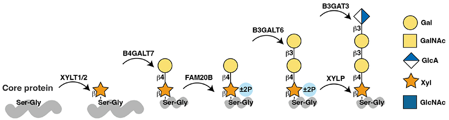
Proteoglycan biosynthesis proceeds through the secretory pathway. First, newly translated core proteins are folded in the lumen of the endoplasmic reticulum and then transported to the Golgi apparatus. In the Golgi lumen, GAG chain synthesis is initiated when XYLT1/2 transfers Xyl from UDP-Xyl to specific Ser residues of core proteins (Figure 2). Among proteins that mature through the Golgi apparatus, approximately 100 are known to become proteoglycans1,5. In many cases, GAG chains are attached to Ser residues within Ser-Gly sequences; however, not all Ser-Gly sequences are modified with GAG chains. Thus, a clear consensus sequence for GAG attachment remains undefined. Recent crystallographic analysis of XYLT1 has provided a structural explanation for its substrate recognition, showing that small amino acids such as Gly or Ala are preferred at the +1 position immediately after the Ser residue and that surrounding acidic residues also contribute to substrate recognition6.
After Xyl transfer, B4GALT7, B3GALT6, and B3GAT3 sequentially add two Gal residues and one GlcA residue, forming the linkage tetrasaccharide GlcAβ1-3Galβ1-3Galβ1-4Xylβ1-O-Ser2,3. It has been reported that the 2-position of Xyl is transiently phosphorylated after addition of the first Gal by B4GALT7. In 2009 FAM20B was identified as the enzyme responsible for this reaction7. Subsequent work showed that FAM20B-mediated Xyl phosphorylation markedly promotes addition of the second Gal by B3GALT6 and functions as a molecular switch that regulates completion of the linkage tetrasaccharide and subsequent GAG chain elongation8. As discussed below, however, this phosphorylation is transient and is removed before polymerization of the GAG chain begins.
In humans, mutations in genes encoding enzymes involved in linkage tetrasaccharide synthesis cause inherited disorders with diverse pathological manifestations. Interestingly, although B3GALT6 deficiency severely impairs GAG synthesis, GAG chains can in some cases be elongated on a noncanonical trisaccharide structure, GlcAβ1-3Galβ1-4Xylβ1-O-Ser9,10.
After completion of the linkage tetrasaccharide, addition of GalNAc initiates CS chain synthesis, whereas addition of GlcNAc initiates HS chain synthesis (Figure 3). Because CS and HS chains share the same linkage tetrasaccharide, the molecular mechanisms that determine whether a given core protein becomes modified with CS or HS have long remained unclear. Early studies using the core protein betaglycan showed that acidic amino acid clusters and Trp residues surrounding Ser-Gly sequences promote HS chain formation11. Recent work has shown that CSGALNACT1/2, the enzymes that initiate CS synthesis by transferring the first GalNAc to the linkage tetrasaccharide, can utilize a relatively broad range of core protein substrates12. In contrast, EXTL3, the HS initiation enzyme that transfers GlcNAc, recognizes acidic sequences in the core protein12. These findings suggest that sequence information within the core protein is important for HS addition. However, a study using thrombomodulin, a CS proteoglycan, also showed that acidic amino acids surrounding the Ser-Gly sequence are important for CS chain elongation13. Thus, many aspects of the sorting mechanism between CS and HS chains remain unresolved.
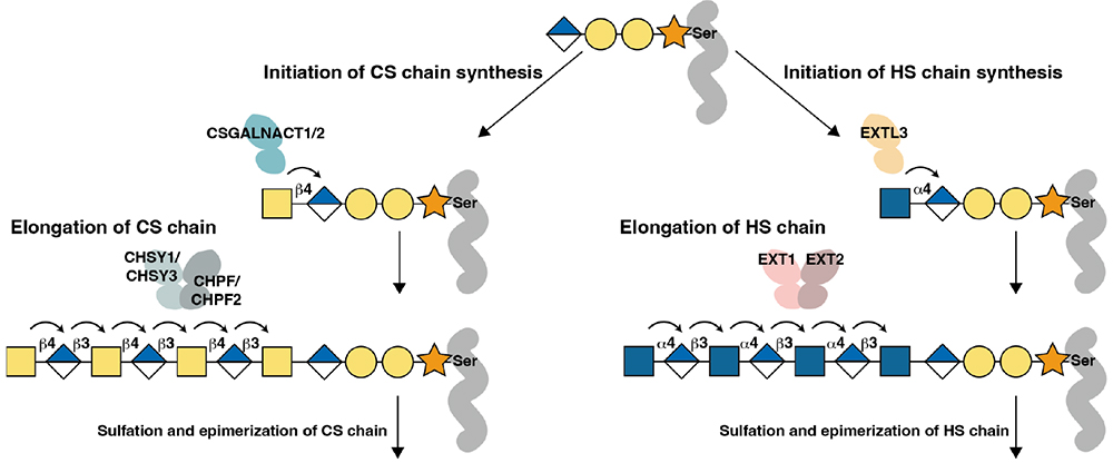
CS chain synthesis is initiated when CSGALNACT1/2 transfers GalNAc to the terminal GlcA of the linkage tetrasaccharide via a β1,4 linkage (Figure 3). CSGALNACT1 preferentially uses the FAM20B-phosphorylated linkage tetrasaccharide as a substrate compared with the nonphosphorylated form14. Subsequent elongation of the repeating disaccharide backbone, [-4GlcAβ1-3GalNAcβ1-]n, involves a group of glycosyltransferases, including CHSY1, CHSY3, CHPF, and CHPF22,15. CHSY1 and CHSY3 possess both GlcA transferase and GalNAc transferase activities within a single enzyme. In contrast, CHPF and CHPF2 were identified as proteins homologous to CHSY1/3, but they do not exhibit clear glycosyltransferase activity by themselves. Nevertheless, CHPF/CHPF2 are required for CHSY1/CHSY3 to elongate repeating disaccharide units. Recent cryo-electron microscopy analysis demonstrated that CHSY1/CHSY3 and CHPF/CHPF2 form functional heterodimers16. This finding indicates that CS chain elongation is regulated by the formation of enzyme complexes. In addition, these polymerase complexes can directly elongate CS chains from the linkage tetrasaccharide even in the absence of CSGALNACT1/22.
Structural diversity in CS chains arises from sulfation of the repeating disaccharide units at various positions. CS sulfation can be broadly divided into two pathways: 4-O-sulfation of GalNAc by C4ST1 (also known as CHST11) and C4ST2 (also known as CHST12), and 6-O-sulfation of GalNAc by C6ST1 (also known as CHST3). Additional sulfation, such as 6-O-sulfation of 4-O-sulfated GalNAc or 2-O-sulfation of GlcA adjacent to 6-O-sulfated GalNAc, generates various sulfated disaccharide structures within CS chains2,4.
Sulfation does not simply occur after completion of the repeating disaccharide backbone; rather, it is thought to occur during CS chain elongation. For example, 4-O-sulfation of GalNAc located at the nonreducing terminus of a growing CS chain promotes CS chain elongation. Therefore, loss of C4ST1 not only abolishes 4-O-sulfated structures but also reduces total CS chain levels2. Recent work has shown that FAM20C interacts with C4ST1 and regulates the sulfation balance of CS chains17. In humans, disruption of this mechanism is thought to cause inherited osteosclerotic bone dysplasia.
During CS chain biosynthesis, some GlcA residues in the repeating disaccharide backbone can be epimerized to IdoA. Subsequent sulfation of GalNAc adjacent to IdoA at the 4-O position by D4ST1 (also known as CHST14) generates dermatan sulfate chains, which exert functions distinct from those of CS chains. Decorin, an important regulator of connective tissue mechanical properties, is a representative proteoglycan bearing dermatan sulfate chains. The importance of dermatan sulfate is further supported by the fact that mutations in D4ST1 or epimerases cause connective tissue fragility disorders18. However, the mechanisms by which only CS chains elongated on specific core proteins are converted into dermatan sulfate chains remain unclear.
HS chain synthesis is initiated by EXTL3-mediated addition of GlcNAc to the linkage tetrasaccharide through an α1,4 linkage3,12 (Figure 3). Recent cryo-electron microscopy analysis revealed that EXTL3 contains two glycosyltransferase domains; one mediates GlcNAc transferase activity, whereas the other has a structure that is poorly suited for glycosyltransferase activity19. Elongation of the HS repeating disaccharide backbone, [-4GlcAβ1-4GlcNAcα1-]n, involves EXT1 and EXT23. Although both EXT1 and EXT2 contain two glycosyltransferase domains, they do not function independently. Instead, they form a functional complex that elongates HS chains3. Recent structural analysis showed that, within the EXT1-EXT2 complex, the GT-B domain of EXT1 corresponds to GlcA transfer, whereas the GT-A domain of EXT2 corresponds to GlcNAc transfer20. These findings indicate that EXTL3 primarily functions in HS chain initiation, whereas the repeating disaccharide backbone of HS is synthesized through coordinated reactions mediated by multiple glycosyltransferase domains within the EXT1-EXT2 complex.
During elongation, HS chains are sequentially modified by various enzymes. First, a subset of GlcNAc residues is N-deacetylated by the NDST family and subsequently N-sulfated. The resulting GlcNS residues serve as starting points for further HS modifications. Recent work has provided a structural basis for the cooperative action of the N-deacetylase and N-sulfotransferase domains of NDST1 in forming N-sulfated regions within HS chains21.
In N-sulfated regions, some GlcA residues are further epimerized to IdoA by GLCE. Subsequently, HS2ST sulfates the 2-O position of IdoA or GlcA, and HS6ST sulfates the 6-O position of GlcNAc or GlcNS. In some regions, HS3ST further sulfates the 3-O position of GlcNS3. Although 3-O-sulfation is a relatively rare modification, it generates structures that are important for recognition by specific proteins such as antithrombin.
A defining feature of HS chains is that these modifications are not uniformly distributed along the entire chain but are concentrated in specific regions. As a result, HS chains contain less sulfated regions as well as highly sulfated domains enriched in N-sulfation, 2-O-sulfation, and 6-O-sulfation22. Through these domain structures, HS chains interact with diverse signaling molecules, including morphogens and growth factors, and regulate cell proliferation, differentiation, morphogenesis, and inflammatory responses.
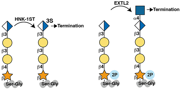
Even for the same core protein, differences in developmental stage or cell type can determine whether GAG chains are elongated to generate proteoglycans or whether GAG chain elongation does not occur. Several mechanisms are known to regulate this switch in GAG chain elongation (Figure 4). First, sulfation of the 3-O position of GlcA located at the nonreducing terminus of the linkage tetrasaccharide by HNK-1ST, also known as CHST10, inhibits subsequent GAG chain elongation23,24. Such sulfated linkage tetrasaccharides are found on aggrecan, a CS proteoglycan expressed in the brain25.
In the normal biosynthetic process, phosphorylated Xyl in the linkage tetrasaccharide is dephosphorylated before polymerization of GAG chains begins. XYLP has been identified as the phosphatase responsible for this reaction26. In contrast, EXTL2 can add GlcNAc through an α1,4 linkage to the phosphorylated linkage tetrasaccharide. The resulting phosphorylated pentasaccharide is not used as a substrate for HS or CS chain elongation, thereby inhibiting subsequent GAG chain elongation27. Indeed, increased production of HS and CS chains has been reported in the liver and brain of EXTL2-deficient mice27.
Another mechanism involves binding of specific proteins to core proteins, which can inhibit GAG chain elongation. For example, a subset of neurexin, a cell adhesion molecule, is modified with HS chains. Binding of its ligand CA10 to neurexin within the secretory pathway suppresses HS addition28. These findings indicate that the use of GAG attachment sites can be regulated not only by core protein sequence, but also by structural modifications of the linkage tetrasaccharide within the secretory pathway and by proteins that bind to core proteins.
Elongation of GAG chains also requires transport of nucleotide sugars, the substrates for glycosyltransferases, from the cytoplasm into the Golgi lumen. SLC35D1 is involved in the transport of UDP-GlcA, UDP-GalNAc, and related nucleotide sugars. Loss of this gene reduces the amount and length of cartilage CS chains in mice and causes chondrodysplasia in humans29. In addition, mice lacking SLC35A3, which is involved in UDP-GlcNAc transport, show reduced CS chains as well as HS chains; however, the mechanism by which CS chains are reduced remains incompletely understood30.
Recent studies have suggested that GAG biosynthetic enzymes are not randomly distributed within the Golgi apparatus, but are instead organized in a manner that allows sequential glycan modification of core proteins22,31,32. For example, super-resolution imaging analysis revealed that the Golgi apparatus is composed of multiple “units” and that each unit contains distinct “zones” of glycosylation enzymes32. The zone of the GAG biosynthetic enzyme XYLT2 differs in localization and dynamics from the zones of the N-glycan enzyme MGAT1 and the mucin-type O-GalNAc glycan enzyme GALNT632. In addition, loss of GRASP55/65, which are required for maintaining Golgi structure, impairs GAG synthesis, sulfation, and secretion33. These findings indicate that the spatial organization of the Golgi apparatus and the dynamics of glycosylation enzymes regulate GAG biosynthesis.
Recent advances in cryo-electron microscopy and machine-learning tools such as AlphaFold have begun to provide structural insights into the catalytic mechanisms of individual enzymes involved in proteoglycan biosynthesis. Nevertheless, many questions remain unresolved. As discussed above, the full mechanism by which CS and HS chain formation is sorted remains unclear. To address this issue, high-resolution analysis of the spatial organization of enzymes involved in CS and HS chain elongation and modification within the Golgi apparatus may be useful. This approach should complement structural studies of individual enzymes. Moreover, recent comprehensive analyses of O-GalNAc glycans have shown that lectican-type CSPGs, including neurocan, brevican, and versican, are enriched not only in CS chains but also in O-GalNAc glycans34. How GAG chains and other glycans are synthesized on proteoglycans in terms of order and subcellular localization also remains an important area for future research. Further advances in glycoproteomics are making it increasingly possible to comprehensively identify which sites on which core proteins are modified with GAG chains5. However, because it remains difficult to analyze intact GAG chains by mass spectrometry, in most cases it is still not possible to determine the length and sulfation pattern of the GAG chain attached to each site. This challenge may be addressed by future advances in mass spectrometry and nanopore-based measurement technologies. A related unresolved issue is how the length of GAG chains, which are synthesized in a template-independent manner unlike DNA and proteins, is controlled. For example, even within the same cell, the length and sulfation pattern of GAG chains can differ depending on the core protein to which they are attached. Elucidating the mechanisms of core protein-selective GAG biosynthesis is therefore an important future challenge.
Proteoglycans are deeply involved not only in development and tissue homeostasis, but also in disease pathogenesis. Therefore, elucidating their biosynthetic mechanisms is expected to contribute not only to a fundamental understanding of glycobiology, but also to clarification of disease mechanisms and the development of new technologies for artificially controlling proteoglycan structures.