Jun 02, 2025

Ubiquitination mediated by sugar
(Glycoforum. 2025 Vol.28 (3), A13)
DOI: https://doi.org/10.32285/glycoforum.28A13

Yukiko Yoshida

吉田 雪子

Yukiko Yoshida
Laboratory Head
Laboratory of Protein Metabolism, Tokyo Metropolitan Institute of Medical Science
She received her Ph.D. in1994 from the Graduate School of Agricultural and Life Sciences at the University of Tokyo, and then worked as a postdoctoral fellow studying glycobiology at RIKEN (Wako). She has been working at the Tokyo Metropolitan Institute of Medical Science since 1997. Her research focus has been on the physiological functions of the ubiquitin system-related glycoproteins.

1. Abstract

The ubiquitin-proteasome system is the major pathway for selective degradation in eukaryotes. Ubiquitin attaches to lysine residues of a substrate, thereby forming ubiquitin chains with various topologies and linkages. Distinct ubiquitin chains serve diverse functions. Recent reports have emerged on the ubiquitination of non-proteinaceous biomolecules, such as lipids and sugars, signifying a rapid expansion of the field of ubiquitination. Here, I introduce the sugar-mediated ubiquitination of Nrf1, a transcription factor of the proteasome, which occurs in peptide: N-glycanase (NGLY1) deficiency, a rare disease.

2. The ubiquitination pathway

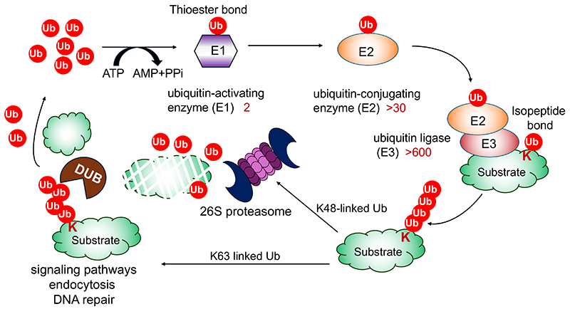
Ubiquitination is mediated by a cascade of three distinct kinds of enzymes: 2 kinds of ubiquitin-activating enzymes (E1s), ~ 30 kinds of ubiquitin-conjugating enzymes (E2), and more than 600 kinds of ubiquitin ligases (E3). Ubiquitination generally involves the formation of isopeptide bonds between the C-terminal glycine of ubiquitin and the ɛ-amino group of the lysine side chains of substrate proteins1-3.

The attached ubiquitin can be further ubiquitinated on its own lysine residues (K6, K11, K27, K29, K33, K48, K63) or the α-amino group of the N-terminal methionine to form polyubiquitin chains. Among them, K48- and K63-linked ubiquitin chains are the most abundant and together account for 70-90% of all linkage types in mammals. K48-linked chains serve as signals for proteasomal degradation, and K63-linked chains act as signals for endocytosis, DNA damage responses, and intracellular signaling events4-6.

The removal of ubiquitin chains is catalyzed by deubiquitinating enzymes (DUBs) following the completion of their respective functions. Consequently, the ubiquitination reaction is a reversible and dynamic process (Fig. 1)5.

図1
Fig.1. The ubiquitination pathway

3. Ubiquity of the ubiquitination signal

Lysine residues are the canonical ubiquitination sites, given the fact that more than 60,000 lysine residues modified with ubiquitin are annotated in the proteome8. However, ubiquitin can be attached to initial methionine (peptide bond), serine or threonine (oxyester bond), or cysteine (thioester bond) residues in proteins9-12.

Furthermore, non-proteinaceous substrates including the lipid A component of lipopolysaccharides (LPS) of Salmonella, phosphatidylethanolamine (PE), ADP-ribose, and glycogen can be ubiquitinated13-17. Although the significance of non-canonical ubiquitination events remains to be fully elucidated, their occurrence is more widespread than currently supposed.

4. Nrf1, a transcription factor for the proteasome

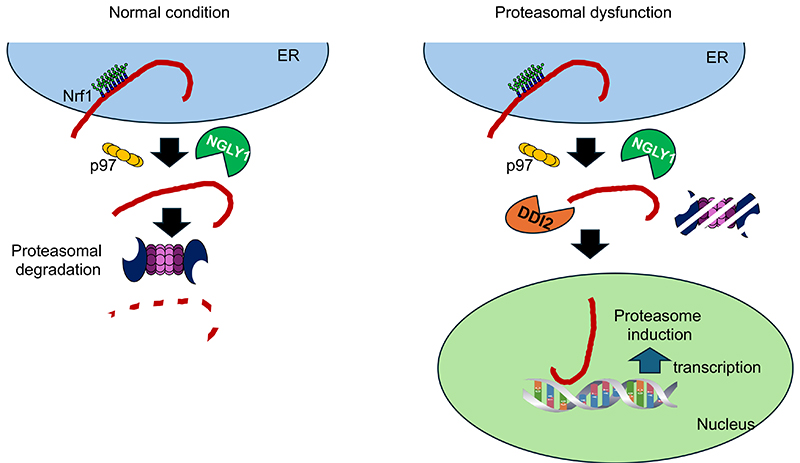
The ubiquitin-proteasome system is essential for cell survival, as it regulates cellular events including the cell cycle and DNA repair18. The proteasome is a large protein complex composed of 66 subunits, and its expression is regulated by the transcription factor Nrf119,20. Nrf1 is synthesized as an N-glycosylated, single-pass membrane protein in the endoplasmic reticulum (ER). In general, misfolded proteins in the ER are retrotranslocated to the cytosol by p97, an AAA-ATPase complex, and degraded by proteasomes, a process called “ER-associated degradation” (ERAD) (Fig. 2). At least under steady state conditions, Nrf1 is constitutively targeted to the ERAD pathway, probably due to its large unstructured domain in the ER, and the amount of the Nrf1 is maintained at low levels in the cells21,22. Under conditions of compromised proteasomal activity, Nrf1 escapes degradation and is transported to the nucleus, where it induces the expression of genes encoding proteasome subunits. In this process, Nrf1 activation requires cleavage by DNA-damage inducible 1 homolog 2 (DDI2) and deglycosylation by peptide: N-glycanase (NGLY1)23-26. Mutations in NGLY1 gene result in NGLY1 deficiency, a rare autosomal recessive congenital disorder, and the expression of the proteasome is reduced in this disease27.

図2
Fig.2. Nrf1 activation pathway under proteasome dysfunction conditions
Nrf1 is N-glycosylated in the endoplasmic reticulum (ER) and extracted from the ER by p97 ATPase. At steady state, Nrf1 is constitutively degraded by the proteasome. Upon proteasomal dysfunction, Nrf1 escapes degradation, is cleaved by the DNA-damage inducible 1 homolog 2 (DDI2), and is deglycosylated by N-glycanase 1 (NGLY1). The activated Nrf1 by DDI2 and NGLY1 is transported into the nucleus, where it activates compensatory proteasome biogenesis.

5. The mysterious connection among cytosolic glycan-related enzymes

As animal models for NGLY1 deficiency, NGLY1 orthologs-deficient models such as C. elegans, Drosophila, mice, and rats, have provided important findings for NGLY1 study (28-30). Notably, the knockout of NGLY1 in Ngly1-KO mice is embryonic lethal on a C57BL/6 genetic background, indicating that NGLY1 activity is essential for their development. Interestingly, the absence of a gene encoding endo-β-N-acetylglucosaminidase (ENGASE), another cytosolic deglycosylating enzyme, partially rescues Ngly1-KO mice from embryonic lethality, yet the double-KO mice show some progressive defects (Fig. 3)31. We observed that additional deletion of the FBS2 gene completely restored the viability of Ngly1-KO mice, and that Ngly1;Fbs2-dKO mice grow without abnormalities. FBS2 is a component of the SKP1-CUL1-F-box (SCF) E3 ubiquitin ligase complex and recognizes N-glycans of glycoproteins32. Therefore, FBS2 ubiquitination activity seems to be the primary cause of the lethality in Ngly1-KO mice. In addition, FBS2 overexpression in NGLY1-KO cells induces cell death, indicating ubiquitination of certain glycoproteins is detrimental to cells lacking NGLY1 activity33.

図3
Fig.3. Three glycan-related enzymes in the cytosol (A) and Ngly1-KO-related mice phenotypes (B)
A. N-glycanase 1 (NGLY1) removes N-glycans from glycoproteins, resulting in the conversion of glycosylated asparagine to aspartic acid. Endo-β-N-acetylglucosaminidase (ENGASE) cleaves between N-acetylglucosamine residues. Skp, Cullin, F-box containing complex (SCFFBS2) ubiquitinates glycoproteins by recognizing N-glycans.
B. The knockout of NGLY1 in Ngly1-KO mice is embryonic lethal on a C57BL/6 genetic background, but additional deletion of ENGase or Fbs2 genes restores viability. The potential glycoforms of cytosolic glycoproteins are indicated.

6. Ubiquitination on the N-acetylglucosamine (GlcNAc) of Nrf1 by FBS2

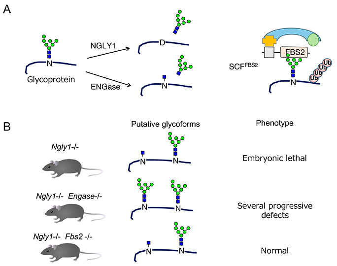
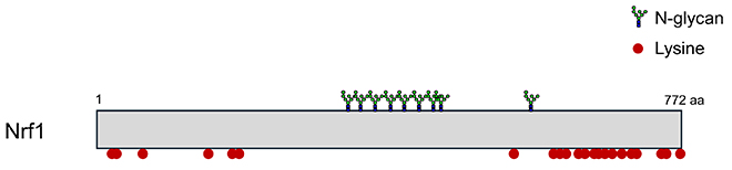
We found that Nrf1 was highly ubiquitinated in FBS2-overexpressing NGLY1-KO cells. However, lysine residues, the usual ubiquitination sites, are not found in Nrf1 around the sites of N-glycosylation recognized by FBS2 (Fig. 4). Furthermore, Nrf1 ubiquitination was not detected in NGLY1:ENGASE-dKO cells, but ubiquitination was restored by expressing ENGASE, indicating that Nrf1 ubiquitination is required for FBS2 and ENGASE activities in the absence of NGLY134. Since ENGASE generates N-GlcNAc on glycoproteins, we hypothesized that SKP1-Cullin1-F-box protein-RBX1 (SCFFBS2) E3 ubiquitin ligase complex could ubiquitinate N-GlcNAc on Nrf1 (Fig. 3A)34.

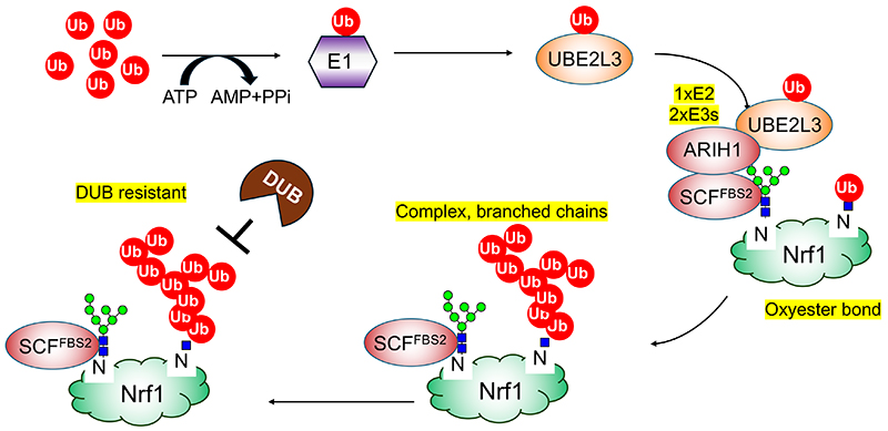
Using an in vitro ubiquitination reconstitution system with chemically synthesized glycopeptides, we found several unique features of this ubiquitination as follows (Fig. 5). First, this ubiquitination occurs through oxyester bonds on serine or threonine residues near N-glycans, as well as the 6-position hydroxy group of N-GlcNAc residues generated from deglycosylated by ENGASE. Second, SCFFBS2 cooperates with another E3 ligase ARIH1. ARIH1 is known to bind to neddylated Cullin-RING E3 ligases (CRLs) and mediate the addition of the first ubiquitin on substrate35,36, but in this case SCFFBS2 captures N-glycans of Nrf1, and ARIH1 ubiquitinates them. Third, the ubiquitin chains are complex, branched, and attached through K6, K11, K33, and K48 ubiquitin linkages. As a result, the atypical ubiquitin chains inactivate Nrf1 by preventing its nuclear transport34.

図4
Fig.4. N-glycosylation sites and positions of lysine residues in Nrf1
図5
Fig. 5. Non-canonical ubiquitination of Nrf1 by N-glycan-recognizing Skp1-Cullin1-F-box-RBX1 containing complex (SCFFBS2)-ARIH1 E3 ubiquitin ligases.

7. Conclusion

I introduced here that ubiquitination also occurs in the glycans of glycoproteins. While the enzymes involved in ubiquitination are located in the cytoplasm and nucleus, the sugar chains are usually located outside the cell or inside the secretory organelles (endoplasmic reticulum, Golgi apparatus, lysosomes, endosomes), and the two are separated by a membrane. Therefore, for a glycoprotein to be ubiquitinated, it must appear in the cytoplasm or in the nucleus (37). In the case of Nrf1, it is thought that this appearance in the cytosol is due to ER-associated degradation, but there are several other possibilities, such as damage to organelles by bacteria or foreign cells that have invaded the body. It will be interesting to see whether such ubiquitination of glycoprotein sugars is seen in other proteins, and whether more non-proteinaceous ubiquitination will be discovered in the future.


References

  1. Hershko A, Ciechanover A. The ubiquitin system. Annu Rev Biochem. 1998;67:425-79.
  2. Pickart CM. Mechanisms underlying ubiquitination. Annu Rev Biochem. 2001;70:503-33.
  3. Deshaies RJ, Joazeiro CA. RING domain E3 ubiquitin ligases. Annu Rev Biochem. 2009;78:399-434.
  4. Kwon YT, Ciechanover A. The Ubiquitin Code in the Ubiquitin-Proteasome System and Autophagy. Trends Biochem Sci. 2017;42(11):873-86.
  5. Swatek KN, Komander D. Ubiquitin modifications. Cell Res. 2016;26(4):399-422.
  6. Komander D, Rape M. The ubiquitin code. Annu Rev Biochem. 2012;81:203-29.
  7. Mevissen TET, Komander D. Mechanisms of Deubiquitinase Specificity and Regulation. Annu Rev Biochem. 2017;86:159-92.
  8. Agrata R, Komander D. Ubiquitin-A structural perspective. Mol Cell. 2025;85(2):323-46.
  9. Cadwell K, Coscoy L. Ubiquitination on nonlysine residues by a viral E3 ubiquitin ligase. Science. 2005;309(5731):127-30.
  10. Shimizu Y, Okuda-Shimizu Y, Hendershot LM. Ubiquitylation of an ERAD substrate occurs on multiple types of amino acids. Mol Cell. 2010;40(6):917-26.
  11. Wang X, Herr RA, Rabelink M, Hoeben RC, Wiertz EJ, Hansen TH. Ube2j2 ubiquitinates hydroxylated amino acids on ER-associated degradation substrates. J Cell Biol. 2009;187(5):655-68.
  12. Kelsall IR, Zhang J, Knebel A, Arthur JSC, Cohen P. The E3 ligase HOIL-1 catalyses ester bond formation between ubiquitin and components of the Myddosome in mammalian cells. Proc Natl Acad Sci U S A. 2019;116(27):13293-8.
  13. Otten EG, Werner E, Crespillo-Casado A, Boyle KB, Dharamdasani V, Pathe C, et al. Ubiquitylation of lipopolysaccharide by RNF213 during bacterial infection. Nature. 2021;594(7861):111-6.
  14. Yang CS, Jividen K, Spencer A, Dworak N, Ni L, Oostdyk LT, et al. Ubiquitin Modification by the E3 Ligase/ADP-Ribosyltransferase Dtx3L/Parp9. Mol Cell. 2017;66(4):503-16 e5.
  15. Chatrin C, Gabrielsen M, Buetow L, Nakasone MA, Ahmed SF, Sumpton D, et al. Structural insights into ADP-ribosylation of ubiquitin by Deltex family E3 ubiquitin ligases. Sci Adv. 2020;6(38):eabc0418.
  16. Kelsall IR, McCrory EH, Xu Y, Scudamore CL, Nanda SK, Mancebo-Gamella P, et al. HOIL-1 ubiquitin ligase activity targets unbranched glucosaccharides and is required to prevent polyglucosan accumulation. EMBO J. 2022;41(8):e109700.
  17. Sakamaki JI, Ode KL, Kurikawa Y, Ueda HR, Yamamoto H, Mizushima N. Ubiquitination of phosphatidylethanolamine in organellar membranes. Mol Cell. 2022;82(19):3677-92 e11.
  18. Collins GA, Goldberg AL. The Logic of the 26S Proteasome. Cell. 2017;169(5):792-806.
  19. Steffen J, Seeger M, Koch A, Krüger E. Proteasomal degradation is transcriptionally controlled by TCF11 via an ERAD-dependent feedback loop. Mol Cell. 2010;40(1):147-58.
  20. Radhakrishnan SK, Lee CS, Young P, Beskow A, Chan JY, Deshaies RJ. Transcription factor Nrf1 mediates the proteasome recovery pathway after proteasome inhibition in mammalian cells. Mol Cell. 2010;38(1):17-28.
  21. Kim HM, Han JW, Chan JY. Nuclear Factor Erythroid-2 Like 1 (NFE2L1): Structure, function and regulation. Gene. 2016;584(1):17-25.
  22. Radhakrishnan SK, den Besten W, Deshaies RJ. p97-dependent retrotranslocation and proteolytic processing govern formation of active Nrf1 upon proteasome inhibition. Elife. 2014;3:e01856.
  23. Koizumi S, Irie T, Hirayama S, Sakurai Y, Yashiroda H, Naguro I, et al. The aspartyl protease DDI2 activates Nrf1 to compensate for proteasome dysfunction. Elife. 2016;5:e18357.
  24. Lehrbach NJ, Ruvkun G. Proteasome dysfunction triggers activation of SKN-1A/Nrf1 by the aspartic protease DDI-1. Elife. 2016;5:e17721.
  25. Tomlin FM, Gerling-Driessen UIM, Liu YC, Flynn RA, Vangala JR, Lentz CS, et al. Inhibition of NGLY1 Inactivates the Transcription Factor Nrf1 and Potentiates Proteasome Inhibitor Cytotoxicity. ACS Cent Sci. 2017;3(11):1143-55.
  26. Lehrbach NJ, Breen PC, Ruvkun G. Protein Sequence Editing of SKN-1A/Nrf1 by Peptide:N-Glycanase Controls Proteasome Gene Expression. Cell. 2019;177(3):737-50 e15.
  27. Rauscher B, Mueller WF, Clauder-Münster S, Jakob P, Islam MS, Sun H, et al. Patient-derived gene and protein expression signatures of NGLY1 deficiency. J Biochem. 2022;171(2):187-99.
  28. Lehrbach NJ. NGLY1: insights from Caenorhabditis elegans. J Biochem. 2022;171(2):145-52.
  29. Pandey A, Jafar-Nejad H. Tracing the NGLY1 footprints: insights from Drosophila. J Biochem. 2022;171(2):153-60.
  30. Fujihira H, Asahina M, Suzuki T. Physiological importance of NGLY1, as revealed by rodent model analyses. J Biochem. 2022;171(2):161-7.
  31. Fujihira H, Masahara-Negishi Y, Tamura M, Huang C, Harada Y, Wakana S, et al. Lethality of mice bearing a knockout of the Ngly1-gene is partially rescued by the additional deletion of the Engase gene. PLoS Genet. 2017;13(4):e1006696.
  32. Yoshida Y, Tokunaga F, Chiba T, Iwai K, Tanaka K, Tai T. Fbs2 is a new member of the E3 ubiquitin ligase family that recognizes sugar chains. J Biol Chem. 2003;278(44):43877-84.
  33. Yoshida Y, Asahina M, Murakami A, Kawawaki J, Yoshida M, Fujinawa R, et al. Loss of peptide:N-glycanase causes proteasome dysfunction mediated by a sugar-recognizing ubiquitin ligase. Proc Natl Acad Sci U S A. 2021;118(27).
  34. Yoshida Y, Takahashi T, Ishii N, Matsuo I, Takahashi S, Inoue H, et al. Sugar-mediated non-canonical ubiquitination impairs Nrf1/NFE2L1 activation. Mol Cell. 2024;84(16):3115-27 e11.
  35. Kelsall IR, Duda DM, Olszewski JL, Hofmann K, Knebel A, Langevin F, et al. TRIAD1 and HHARI bind to and are activated by distinct neddylated Cullin-RING ligase complexes. EMBO J. 2013;32(21):2848-60.
  36. Scott DC, Rhee DY, Duda DM, Kelsall IR, Olszewski JL, Paulo JA, et al. Two Distinct Types of E3 Ligases Work in Unison to Regulate Substrate Ubiquitylation. Cell. 2016;166(5):1198-214 e24.
  37. Yoshida Y, Tanaka K. Cytosolic N-Glycans: Triggers for Ubiquitination Directing Proteasomal and Autophagic Degradation: Molecular Systems for Monitoring Cytosolic N-Glycans as Signals for Unwanted Proteins and Organelles. Bioessays. 2018;40(3):1700215.
top