PTP

is a receptor-type protein tyrosine phosphatase, which is synthesized as a chondroitin sulfate proteoglycan. The extracellular region of this receptor is secreted as a soluble brain-specific chondroitin sulfate proteoglycan, phosphacan. PTP
uses a heparin-binding growth factor, pleiotrophin, as a ligand. The chondroitin sulfate portion of this receptor is essential for binding to pleiotrophin, and it is conceivable that D unit [GlcUA(2S)-GalNAc(6S)]-containing chondroitin sulfate chains are involved in the high affinity binding and the signal transduction (1). Recently, we found that PTP
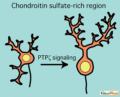
-pleiotrophin signaling regulates the morphogenesis of cerebellar Purkinje cells (2). By the inhibition of this signaling system, an aberrant morphology of Purkinje cell dendrites such as multiple and disoriented primary dendrites was induced in the organotypic slice cultures of rat cerebellum (
Fig. A). It is remarkable that Purkinje cells are surrounded by the D unit-rich chondroitin sulfate, which is involved in the binding to pleiotrophin.
Sugahara et al. (3) revealed that oversulfated chondroitin sulfate and dermatan sulfate promote the neurite outgrowth of hippocampal neurons form embryonic mouse. Hippocampal neurons cultured on substrates coated with D or iD unit [GlcUA/IdoUA(2S)-GalNAc(6S)]-rich chondroitin sulfate or dermatan sulfate developed multiple dendrite-like processes. On the other hand, E or iE unit [GlcUA/IdoUA-GalNAc(4S, 6S)]-rich chondroitin sulfate or dermatan sulfate induced the outgrowth of long axon-like neurites. It is known that the oversulfated chondroitin sulfate binds with many heparin-binding growth factors, which may play roles in the neuritogenesis of hippocampal neurons.
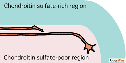
In the above examples, chondroitin sulfate is presented to the neurons as a uniform environmental substance. In the developing nervous system, however, chondroitin sulfate is often distributed discontinuously at some boundary regions. It is considered that such chondroitin sulfate-rich zones inhibit the penetration of growing axons, guiding the axonal pathways (
Fig. B). Mouse optic chiasm and chick diencephalo-telencepalic boundary are good examples, and chondroitinase ABC digestion of these regions disrupted the navigation of retinal axons (4). Recent investigations revealed that not only the differences in concentration but also the structural variation of chondroitin sulfate at decision points control the axon guidance (4). Elucidation of the molecular mechanism of such repulsive effects of chondroitin sulfate will have large medical impact because chondroitin sulfate is also regarded as an inhibitor of axonal regeneration.