| Neuroglycan C (NGC) and Some Other Typical Part-time Proteoglycans | |
|
 |
Proteoglycans are proteins bearing one or more sulfated glycosaminoglycan side chains. Recent studies have indicated that, although many proteins occur solely in a proteoglycan form, certain proteins occur both in a proteoglycan form and in a non-proteoglycan form without sulfated glycosaminoglycans. These proteins are called part-time proteoglycans.
One of the typical part-time proteoglycans is thrombomodulin, a critical mediator of endothelial anticoagulant defenses. Comparative studies on the anticoagulant effects between the proteoglycan form with chondroitin sulfate (ß -TM) and the non-proteoglycan form (a-TM) of thrombomodulin revealed that the chondroitin sulfate moiety plays an important role in regulating the anticoagulation functions. Sugar analysis of a-TM expressed in CHO-K1 cells showed that an oligosaccharide with a structure of GlcA ß1-3Gal ß1-3Gal ß1-4Xyl was liberated from a-TM by mild alkaline treatment (1). This oligosaccharide represents a glycosaminoglycan-protein linkage tetrasaccharide common to various proteoglycans, suggesting that the critical determining step for biosynthesis of chondroitin sulfate on thrombomodulin is the transfer of the first N-acetylgalactosamine to produce the repeating disaccharide structure of chondroitin sulfate, rather than the transfer of xylose.
A certain population of Alzheimer amyloid precursor protein (APP) occurs in a molecular structure bearing chondroitin sulfate side chains, which is called appican. This population appears augmented after brain damage induced by excitotoxicity. Interestingly, the core protein of appican derives from an APP mRNA lacking exon 15. Splicing out of this exon creates a new consensus sequence for the transfer of xylose (2). Since the chondroitin sulfate chain is attached to a serine residue close to (16 amino acids upstream of) the amino terminus of the A ß sequence of APP, this attachment could affect the proteolytic processing of APP and production of A ß peptides.
Another interesting example of part-time proteoglycan is neuroglycan C (NGC), a transmembrane glycoprotein with a single EGF module whose expression is restricted in the central nervous system. NGC exists in a proteoglycan form with a single chondroitin sulfate chain in the developing cerebellum of the mouse, whereas it exists largely in a non-proteoglycan form in the mature cerebellum (3). A similar developmental change in the structure of NGC occurs in the rat retina. This is the first example of part-time proteoglycans that changes its molecular structure from a proteoglycan form to a non-proteoglycan form, or vice versa, during tissue development. Immunohistochemical studies showed that only Purkinje cells are immunoreactive with anti-NGC antibodies in the cerebellum. The climbing and parallel fiber systems are the major afferent neural fiber systems to the Purkinje cells. The spatiotemporal expression pattern of NGC on the differentiating Purkinje cells coincides with the pattern of synaptogenesis of the climbing fibers, not the parallel fibers, with the Purkinje cells during cerebellar development. This suggests that the proteoglycan form of NGC in the developing cerebellum mediates adhesion and synaptogenesis of the climbing fibers to the Purkinje cells, and that it may inhibit adhesion of the parallel fibers to the Purkinje cells.
In the extracellular domain of NGC, there are 6 Ser-Gly sequences conserved among mouse, rat and human. However, site-directed mutagenesis showed that only one site is substituted by chondroitin sulfate. No splicing variants of NGC with a peptide deletion to form the chondroitin sulfate attachment site, as seen in the case of appican, have been detected.
It remains to be examined how the synthesis of the chondroitin sulfate chain on the NGC core protein is regulated, and how functional roles of NGC change along with the developmental change in its structure.
| |
|
 |
 |
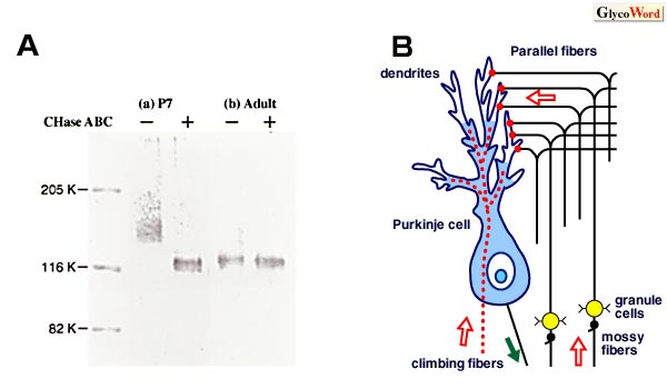
Western blot analysis for NGC in the cerebellum of 7-day-old (P7; a) and adult (b) mice. Tissue homogenates were separated by SDS-PAGE before (-) and after (+) digestion with chondroitinase ABC (CHase ABC), and NGC was visualized by staining with an anti-NGC antibody after proteins were electrotransferred to a PVDF membrane. NGC in the P7 sample was obtained as a broad band around a molecular weight of 150 K. CHase ABC-treatment converted the broad band to a narrow band at 120 K, indicating that NGC occurs in a proteoglycan form with chondroitin sulfate in the developing cerebellum. In the adult sample, a significant amount of the 120 K band was obtained even before digestion with CHase ABC. This indicates that NGC largely exists in a non-proteoglycan form in the adult cerebellum.
|
|
Two major afferent fibers to the Purkinje cells of the cerebellum. The climbing fibers originating from the inferior olivary complex form synapses with thick stems of the Purkinje cell dendrites, whereas the parallel fibers which are the axons of the cerebellar granule cells form synapses with thin branchlets. NGC is localized to the soma and thick stems of the dendrites of the Purkinje cells in the developing cerebellum. |
|
|
|
|
| Atsuhiko Oohira (Institute for Developmental Research, Aichi Human Service Center) | |
|
|
| References | (1) | Nadanaka S , Kitagawa H, Sugahara K : Demonstration of the immature glycosaminoglycan tetrasaccharide sequence GlcA ß1-3Gal ß1-3Gal ß1-4Xyl on recombinant soluble humann a-thrombomodulin. J. Biol. Chem. 273, 33728-33734, 1998
|
|
(2) |
Pangalos MN, Efthimiopoulos S, Shioi J, Robakis NK :The chondroitin sulfate attachment site of appican is formed by splicing out exon 15 of the amyloid precursor gene. J. Biol. Chem. 270, 10388-10391, 1995
|
|
(3) |
Aono S , Keino H , Ono T , Yasuda Y , Tokita Y , Matsui F ,Taniguchi M , Sonta S, Oohira A : Genomic organization and expression pattern of mouse neuroglycan C in the cerebellar development. J. Biol. Chem. 275, 337-342, 2000
|
| |
|
|
|
| Jun. 15, 2001 |
|
| |
|
|
|
|


