 |
Among C-type lectins in marine invertebrates, three-dimensional structures of the tunicate (Polyandrocarpa misakiensis) lectin TC14 (1) and the sea cucumber (Cucumaria echinata) lectin CEL-I (2) were determined by X-ray crystallographic analyses. They are composed of two C-type carbohydrate-recognition domains (CRDs), whose main-chain structures are very similar to those of mannose-binding lectins (MBLs) in mammals. In addition, these lectins bind specific carbohydrates through coordinate bonds with Ca2+ ions and hydrogen bond networks with nearby amino acid residues in a manner which also resembles the binding mode of MBLs. C-type CRDs generally contain Gln-Pro-Asp (QPD) of Glu-Pro-Asn (EPN) motifs, which are assumed to be important for galactose or mannose specificities, respectively. In fact, GalNAc/Gal-specific CEL-I contains a QPD sequence in its carbohydrate-binding site. On the other hand, TC14 contains a Glu-Pro-Ser (EPS) sequence that is similar to the EPN motif, although this lectin shows galactose specificity. This is due to the different orientation of 3-OH and 4-OH groups of bound galactose compared with other C-type CRDs, and stacking between tryptophan side chain and the hydrophobic face of galactose. CEL-I shows very high specificity for N-acetylgalactosamine (GalNAc), which is caused by hydrogen bonds and van der Waals contacts between the acetamido group of GalNAc and the binding site of the lectin. These observations suggest that marine invertebrate C-type lectins may generate diversity in carbohydrate-binding specificity by changing various amino acid residues in the carbohydrate-binding sites, although their basic structures are very similar to those of mammals.
Besides C-type lectin CEL-I, there is another lectin CEL-III, which shows hemolytic as well as cytotoxic activities by damaging target cell membranes after binding to the carbohydrate chains. The three-dimensional structure of CEL-III was also determined by X-ray crystallographic analysis and found to be composed of two N-terminal carbohydrate-binding domains (domains 1 and 2), and a C-terminal oligomerization domain (domain 3) (3). Domains 1 and 2 are composed of ricin-type (R-type) CRDs, which have a  -trefoil structure with pseudo three-fold symmetry. However, the carbohydrate-binding mode of domains 1 and 2 is rather similar to that of C-type CRD because the OH groups of bound carbohydrates are coordinated with Ca2+ and also hydrogen-bonded with nearby amino acid residues in the binding sites. Thus, CEL-III has a unique feature sharing similarities to C-type and R-type CRDs. On the other hand, domain 3 contains extended -sheets and a hydrophobic region, suggesting that this domain plays an important role in self-oligomerization in target cell membranes. CEL-III presumably undergoes conformational change after binding to the cell surface carbohydrate chains, leading to pore-formation in the target membrane. This suggests a mechanism similar to that of bacterial pore-forming toxins. -trefoil structure with pseudo three-fold symmetry. However, the carbohydrate-binding mode of domains 1 and 2 is rather similar to that of C-type CRD because the OH groups of bound carbohydrates are coordinated with Ca2+ and also hydrogen-bonded with nearby amino acid residues in the binding sites. Thus, CEL-III has a unique feature sharing similarities to C-type and R-type CRDs. On the other hand, domain 3 contains extended -sheets and a hydrophobic region, suggesting that this domain plays an important role in self-oligomerization in target cell membranes. CEL-III presumably undergoes conformational change after binding to the cell surface carbohydrate chains, leading to pore-formation in the target membrane. This suggests a mechanism similar to that of bacterial pore-forming toxins. | |
|
| References | (1) |
Poget, S.F., Legge, G.B., Proctor, M.R., Butler, P.J., Bycroft, M. and Williams, R.L. (1999) The structure of a tunicate C-type lectin from Polyandrocarpa misakiensis complexed with D-galactose. J. Mol. Biol.290, 867-879.
|
| (2) |
Sugawara, H., Kusunoki, M., Kurisu, G., Fujimoto, T., Aoyagi, H. and Hatakeyama, T. (2004) Characteristic recognition of N-acetylgalactosamine by an invertebrate C-type lectin, CEL-I, revealed by X-ray crystallographic analysis. J. Biol. Chem. 279, 45219-45225.
|
| (3) |
Uchida, T., Yamasaki, T., Eto, S., Sugawara, H., Kurisu, G., Nakagawa, A., Kusunoki, M. and Hatakeyama, T. (2004) Crystal structure of the hemolytic lectin CEL-III isolated from the marine invertebrate Cucumaria echinata: implications of domain structure for its membrane pore-formation mechanism. J. Biol. Chem.279, 37133-37141.
|
| | |
| |


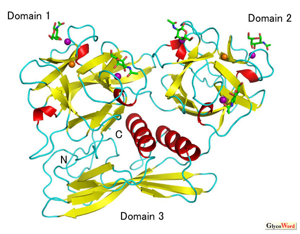
 -sheets are indicated in red and yellow, respectively. Ca2+ and Mg2+ ions are shown as purple and orange balls. There are two and three GalNAc molecules bound in domain 1 and domain 2, respectively.