


Extracellular vesicles and gangliosides | |||||||||||||||
 |
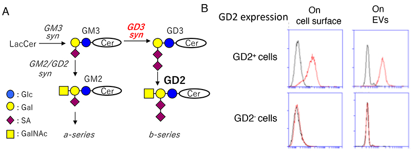
Among extracellular vesicles (EVs), endosome-derived exosomes are different from plasma membrane-derived ectosomes or apoptotic vesicles in the basic properties and functions, and their biological and medical significances are attracting attention (1). Exosomes are 30-160 nm in diameter small vesicles consisting of 2-layer lipid membrane, and secreted from various organisms from bacteria to mammals, containing nucleic acids (DNA, mRNA, microRNA), proteins, and lipids. They play various roles for exchange of cell-to-cell information. However, information on carbohydrates, in particular of glycosphingolipids, in exosomes have been very poor, and their roles, even their presence has scarcely been reported. Here, characters and roles of glycosphingolipids in exosomes (hereafter EVs) secreted from mainly human and mouse cultured cells are introduced mainly with findings in our research group. As for preparation methods of EVs, ultracentrifugation approaches are generally used, and obtained particles seem to be homogenous in size and morphology, proposing no particular issues. But, this method needs long time such as repeated ultracentrifugation, therefore, various alternative and simple methods have been proposed. What is attracting attention regarding EVs are infection-immunity, regulation of differentiation, and cancers, and particularly in cancer studies, usefulness and significance of EVs released from cancer cells in diagnosis and their functions in the regulation of cancer-microenvironment and metastasis. Regarding functions of EVs released from mesenchymal stem-cells in the regulation of tissue differentiation/inflammation, detailed analyses have been performed, and their application for clinical therapeutics is being expected. As for regulatory functions of EVs for cancer-microenvironment, a number of effects on endothelial cells, fibroblasts, immune cells have been reported, suggesting that they are important to generate advantageous environment for cancers. On the other hand, many reports indicate that EVs generally exert to make better conditions for distant metastasis of cancers. In particular, the study that reported site-specific niche formation for metastasis depending on the integrin isoforms in EVs is highly attractive. Actually, EVs secreted from breast cancer cells expressing different isoforms of integrins exert to form niches for organo-tropic metastasis, such as lung, bone, and liver etc (2). Furthermore, composition of miRNAs contained in EVs collected from body liquids such as blood of various cancer patients revealed characteristic composition of miRNA depending on the kinds of cancers, suggesting that EVs can be useful probes for the cancer diagnosis (2). Gangliosides are sialic acid-containing glycosphingolipids, and disialyl gangliosides have been, in particular, used in the cancer diagnosis and therapy as cancer-associated antigens for neuroectoderm-derived cancers (3). Using melanoma and glioma cells, it has been reported that expression of disialyl gangliosides enhances malignant properties and cell signals in various cancer cell systems. Furthermore, many novel findings on the properties and functions of EVs released from cancer-associated ganglioside-expressing cancer cells have been elucidated (Fig. 1).
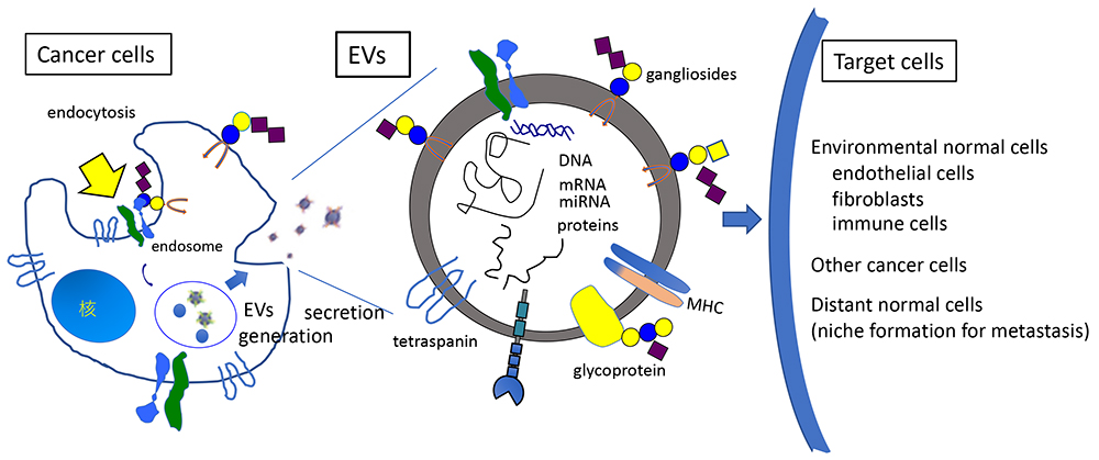
When compositions of EVs were compared with those of cells, profiles of nucleic acids and proteins were largely different, and they were also different depending on the sugar structures in glycolipids expressed on cancer cells. On the other hand, expression and composition of lipids, particularly of glycosphingolipids showed similar patterns between EVs and cells. By establishing melanoma cell lines expressing characteristically different gangliosides from a single parent line, we analyzed secreted EVs from them, showing clearly different functions depending on the expressed glycosphingolipids. For example, EVs released from ganglioside GD2-expressing melanoma cells enhanced malignant properties such as cell growth, invasion, motility and adhesion in GD2-negative melanoma cells in a dose-dependent manner (4). Almost similarly, enhanced malignant properties could be found in glioma cells when EVs from GD3/GD2-expressing cells were added to GD3/GD2-negative glioma cells (5). Moreover, when cell signals were analyzed by immunoblotting after addition of EVs, it was shown that activation of growth factor receptors and adhesion receptors, and their downstream signaling molecules was induced within 15 min. Furthermore, these effects of EVs were suppressed by the addition of anti-ganglioside antibodies, suggesting that gangliosides expressed on the surface of EVs are essentially involved in the action of EVs towards target cells. Interestingly, when EVs from disialyl ganglioside-negative cancer cells were conversely added to -positive cells resulted in the suppression of cell phenotypes and signals. These results suggest that EVs are playing roles in the equalization of heterogenous cancer tissues, leading to the formation of a more malignant and uniform cancer cell group via cell-to-cell communication. In summary, following issues should be main topics of gangliosides in EVs: How does expression of gangliosides affect generation and secretion of EVs in cancer cells? How do EVs access to, invade in, and exert roles in target cells? How is the collaboration dynamics of gangliosides with membrane molecules on the surface of EVs? and How is possibility about therapeutic application of EVs (Fig. 2) are being asked.
Koichi Furukawa / Yuhsuke Ohmi / Keiko Furukawa
| ||||||||||||||
| References | |
|---|---|
| (1) | Urabe F, Kosaka N, Kimura T, Egawa S, Ochiya T: Extracellular vesicles: Toward a clinical application in urological cancer treatment. Int. J. Urol. 25, 533-543, 2018 |
| (2) | Hoshino A, Costa-Silva B, Shen TL, Rodrigues G, Hashimoto A, Tesic Mark M, Molina H, Kohsaka S, Di Giannatale A, Ceder S, Singh S, Williams C, Soplop N, Uryu K, Pharmer L, King T, Bojmar L, Davies AE, Ararso Y, Zhang T, Zhang H, Hernandez J, Weiss JM, Dumont-Cole VD, Kramer K, Wexler LH, Narendran A, Schwartz GK, Healey JH, Sandstrom P, Labori KJ, Kure EH, Grandgenett PM, Hollingsworth MA, de Sousa M, Kaur S, Jain M, Mallya K, Batra SK, Jarnagin WR, Brady MS, Fodstad O, Muller V, Pantel K, Minn AJ, Bissell MJ, Garcia BA, Kang Y, Rajasekhar VK, Ghajar CM, Matei I, Peinado H, Bromberg J, Lyden D: Tumour exosome integrins determine organotropic metastasis. Nature 527, 329-335, 2015. |
| (3) | Furukawa K, Ohmi Y, Hamamura K, Kondo Y, Ohkawa Y, Kaneko K, Hashimoto N, Farhana Y, Bhuiyan RH, Tajima O, Furukawa K: Signaling domains of cancer-associated glycolipids. Tribute to Professor Sen-itiroh Hakomori. Glycoconj. J. 39, 145-155, 2022 |
| (4) | Yesmin F, Furukawa K, Kambe M, Ohmi Y, Bhuiyan RH, Hasnat MA, Mizutani M, Tajima O, Hashimoto N, Kaneko K, Furukawa K: Extracellular vesicles released from ganglioside GD2-expressing melanoma cells enhance malignant properties of GD2-negative melanomas. Sci. Rep. 13, 4987, 2023. |
| (5) | Hasnat MA, Ohmi Y, Yesmin F, Kambe M, Kawamoto Y, Bhuiyan RH, Mizutani M, Hashimoto N, Tsuchida A, Ohkawa Y, Kaneko K, Tajima O, Furukawa K, Furukawa K: Crucial roles of exosomes secreted from ganglioside GD3/GD2-positive glioma cells in enhancement of the malignant phenotypes and signals of GD3/GD2-negative glioma. Nagoya J. Med. Sci. 2024 in press |
Jul. 17, 2024